- Home
- UFAI in the News
- UFAI Medical Journals
- Arthroscopic Approach to Posterior Ankle Impingement
Arthroscopic Approach to Posterior Ankle Impingement
Written by: Dr. Michael H. Theodoulou, DPM and Dr. Laura Bohman, DPM
INTRODUCTION
Posterior impingement syndrome is often synonymous with the terms posterior talar compression syndrome, os trigonum syndrome, posterior ankle block, nutcracker- type impingement, and posterior tibiotalar impingement syndrome. Frequently, it is associated with the presence of an accessory ossicle known as the os trigonum. Pain can be elicited acutely with a forced plantarflexion injury or chronically in those individuals performing repetitive plantarflexory moments of the foot and ankle, such as ballerinas or soccer players. The latter presentation is more common.
The os trigonum develops as a secondary cartilaginous center during the second month of fetal development, between the ages of 7 and 11 years in girls and 11 and 13 years in boys. Through enchondral ossification, the center fuses to the postero- lateral talus with a cartilaginous synchondrosis. One year after its appearance, the secondary center unites to the talar body producing the Stieda process. Failure to unite has been identified in 1.7% to 49%3 of the general population. Intimate to the presence of this accessory ossicle, is the flexor hallucis longus (FHL) tendon. As a result, the tendon is susceptible to a stenosis tenosynovitis.
Chronic entrapment of this tendon can occur resulting from low-lying muscle tissue, impingement from an os trigonum, and incongruity of maximum plantarflexion and dorsiflexion of the ankle and great toe joint resulting in compression of the tendon. This condition often occurs in the fibro-osseous tunnel posterior to the medial malleolus. This condition can be seen in athletes who require forceful plantarflexion of the foot, such as soccer players, swimmers, and ice skaters.
ANATOMY
The posterior ankle joint complex is well defined by soft tissue and osseous structures. Medially, it is bounded by the flexor tendons of the leg, including (from superficial to deep) the posterior tibial tendon, the flexor digitorum longus, and the FHL. The FHL tendon is a critical medial boundary during arthroscopy because the neurovascular bundle lies just medial or posterior to this structure. Posteriorly, the prominent Achilles tendon is appreciated. Laterally, the peroneal tendons serve as an outside boundary to protect the sural nerve and small saphenous vein during arthroscopy. Anteriorly lies the tibiotalar and talocalcaneal joints. Inferiorly, is the tuber of the calca- neus. Within the contents of this vault is adipose tissue frequently refer to as the Kager triangle.
In 2002, Sitler and colleagues published an anatomic study regarding structures at risk when performing posterior ankle arthroscopy through both medial and lateral por- tals. They examined 13 fresh frozen cadavers and placed plastic cannulas filled with oil in portals to serve as landmarks when performing MRI. Imaging studies were compared with open dissection of the specimens to confirm correlation of proximity of vital structures. Portals were placed immediately adjacent to the Achilles tendon. It was appreciated that, on average, the sural nerve was 3.2 mm from the portal, 4.8 mm to the small saphenous vein, 6.4 mm to the tibial nerve, 9.6 mm to the posterior tibial artery, 17 mm to the medial calcaneal nerve, and 2.7 mm to the FHL tendon. There was little discrepancy with MRI studies with the exception of the tibial nerve, which could not always be appreciated on MRI.
Balci and colleagues similarly studied cadaveric specimens, placing posteromedial and lateral portals but adjusting the position of the ankle, assessing neutral, 150 of dor- siflexion, and 300 of plantarflexion. In neutral, the anterolateral portal was 6 mm from the sural nerve and 1.6 mm from the peroneals. The distance between the medial por- tal and the FHL was 2.11 mm and from the tibial artery was 6 mm. With increased dor- siflexion, the distance between the posterior portal and the neurovascular bundle medially increased. This finding may suggest that dorsiflexion of the ankle during por- tal placement may better protect medial vital structures.
PATHOGENESIS
Although a rare condition, os trigonum syndrome may occur acutely through hyper- plantarflexion injury or chronically by repetitive plantarflexion stress moments. As the talus rotates plantarly, the accessory ossicle or prominent lateral talar process is impinged between the calcaneus inferiorly and the tibial plafond superiorly. It must also be appreciated that hyperdorsiflexion of the foot can produce avulsion of the lateral talar process by increased tension to the posterior talofibular ligament. This condition has also been referred to as a Shepherd fracture.
In overuse or chronic repetitive plantarflexion injuries, it is thought that the soft tissue is repetitively impinged between the bone entities producing inflammation that gradually results in changes to the adjacent FHL. Recalling the Balci and col- leagues study, increased plantarflexion of the ankle produces lateralization of the posterior structures, bringing the FHL tendon into greater risk for impingement. In FHL tenosynovitis, patients may report tenderness in the great toe and posteromedial ankle joint.
PATIENT HISTORY AND PHYSICAL FINDINGS
Appropriate history and physical is obtained to establish the nature, location, onset, duration, and aggravating and mitigating influences of the clinical complaint. Under- standing the activities of the individual is also important. In the acute injury, a forced hyperplantarflexion is usually described. In chronic cases of overuse and os trigonum syndrome, pain is frequently noted to the posterolateral aspect of the ankle but can also be seen posteromedial and diffusely to the posterior aspect of the ankle joint.
During the physical examination, palpation across the posterolateral ankle joint is critical to establish trigger point tenderness. There may be focal swelling in this region. Forced plantarflexion of the foot can reproduce impingement and pain to the area, known as the nutcracker sign.
There may be a decrease in ankle range of motion, particularly in plantarflexion. In chronic cases in which the FHL becomes inflamed, reduction of hallux motion can occur secondary to fibrosis. A provocative maneuver for pain reproduction can be done with the knee in extension, dorsiflexion of the ankle, and attempted hallux dorsiflexion.
Diagnostic injections of local anesthetic with image guidance such as fluoroscopy or ultrasonography can help confirm diagnosis if pain relief is achieved with use. Differential diagnosis based on these findings can include a host of conditions to include fractures of the posterior tibiotalocalcaneal complex, osteochondral lesions of the talus and/or tibia, tarsal coalition, soft tissue lesions, and tendon disorders in- clusive of the peroneals and Achilles.
IMAGING
Lateral plain film radiographs are usually adequate to identify the presence of postero- lateral talar prominence or an accessory ossicle. Lateral foot plantarflexion images can show posterior impingement on the distal tibia.. Advanced imaging with computed tomography (CT) and MRI can be used if necessary. CT scans can more accurately establish the relationship of bone structures producing the image. They can also benefit the identification of fracture. MRI can identify marrow edema, fluid collections, and most importantly concomi- tant synovitis of the FHL tendon. Sonography can dynamically examine the gliding of the FHL tendon during passive dorsiflexing and plantarflexing of the ankle.
TREATMENT
As in any inflammatory process producing local pain and dysfunction, initial nonoperative care includes rest, ice, antiinflammatory medication, and avoidance of activities that exacerbate the condition. A period of immobilization in a walking boot can be considered.
Physical therapy to assist in reduction of inflammation may be instituted with progressive range of motion, strengthening, and reconditioning to sport-specific activity. As previously discussed, diagnostic and therapeutic injections of local anesthesia can be administered. If a corticosteroid is used, it should be done with caution to avoid injecting intratendinously. Image guidance using fluoroscopy or ultrasonography can aid with administration. These injections can assist in understanding the response to surgical intervention if considered.
Failure to respond to nonoperative care for a period of 3 to 6 months suggests that oper- ative intervention may be an acceptable alternative. Older literature presents an open technique both through a posteromedial and posterolateral approach.10 With advance- ment of endoscopic technique, much of recent literature is devoted to this approach.
SURGICAL TECHNIQUE
Historically, posterior ankle arthroscopy and/or endoscopy was considered potentially dangerous because of the proximity of vital neurovascular structures. In 2000, van Dijk and colleagues published their approach to posterior ankle arthroendoscopy, creating a method that carefully and systematically avoided the neurovascular bundle. Since that time, posterior impingement treated surgically by an endoscopic approach has been performed more consistently.
Once the patient has completed a 3-month to 6-month course of failed conservative treatment, surgical intervention may be indicated. A thorough preoperative medical work-up should be completed to clear the patient for elective surgery. It is important to take into consideration that the patient will be positioned prone when reviewing pertinent medical history.
POSITIONING
The patient is placed prone on the operating room table with a well-padded thigh or high calf tourniquet. The operative limb is elevated on several blankets, a pillow, or a ramp to raise the affected extremity above the contralateral foot. This elevation prevents restriction of movement for the arthroscopic equipment during the procedure. The pelvis should be bumped accordingly to allow the foot to be posi- tioned perpendicular to the floor with the foot resting in a neutral 900 with respect to the leg.
Anatomic structures are then drawn out. From the tip of the fibula, a line par- allel with the plantar foot is drawn to the medial side. The medial and lateral borders of the Achilles tendon are then marked. The medial and lateral portals are drawn just above the intersection where the medial and lateral borders of the Achilles tendon meet the transverse line. A line is then drawn across the bottom of the foot extending from the lateral portal to the first web space.
TECHNIQUE
The extremity is then scrubbed, prepped, and draped with aseptic technique. The skin overlying the lateral portal is sharply created with care to not penetrate through sub- cutaneous tissue. Blunt dissection using a mosquito hemostat is taken down to the level of bone in the same direction as the line drawn to the first web space. The hemo- stat is then replaced for a 300 4.0 mm endoscope with saline set to gravity (Video 1). The medial portal is then created just medial to the Achilles tendon in the same plane as the lateral portal. A hemostat is inserted through the portal at 900 to the scope just deep to the Achilles tendon. The scope is held pointing to the first web space and is used as a guide for the hemostat to travel down to the level of bone, thereby avoiding the neurovascular bundle. The hemostat is then replaced with a shaver. To obtain a visual of the surrounding structures, some of the fat surrounding the scope must be excised.
Anatomic landmarks and disorders can then be evaluated. The FHL tendon is medial to the Stieda process along the posterior joint capsule. It is important to identify the FHL tendon and work laterally to avoid compromise of the neurovascular structures.
Flexion and extension of the toe aid in this visualization. Ligamentous structures, including the posterior inferior tibiofibular ligament, posterior talofibular ligament, and calcaneofibular ligament, should be visible lateral to the FHL. Deep to the capsule, the posterior aspects of the subtalar and ankle joints can be inspected. These struc- tures are flanked laterally by the fibula and peroneal tendons and medially by the FHL tendon, the neurovascular bundle, the flexor digitorum longus, posterior tibial tendon, and the medial malleolus.
Many disorders can be addressed using this technique, such as posterior ankle and subtalar joint osteochondral lesions, subtalar/ankle arthritis, soft tissue impingement, bony impingement, arthroscopic evaluation of fracture reduction, FHL or peroneal tenosynovitis, and excision of talocalcaneal coalitions (Video 2). The portals can then be closed primarily with application of a soft dressing, splint, or cast.
Alternatively, Horibe and colleagues described a different method for excision of a symptomatic os trigonum. They proposed using the posterolateral portal as described by van Dijk and colleagues, but instead of the typical posterior medial portal they used an accessory posterolateral portal. In the transverse plane, this portal was placed just below the fibular tip and posterior to the peroneal tendons.
REHABILITATION AND RECOVERY
The postoperative protocol differs with each type of procedure. van Dijk and col- leagues placed a soft postoperative dressing after a symptomatic os trigonum exci- sion in a ballet dancer. The patient was then instructed to perform ankle joint range of motion exercises starting immediately after surgery followed by weight bearing after 3 days. Physical therapy was initiated 2 weeks postoperatively and the patient returned to professional dancing after 6 weeks. Gasparetto and colleagues recom- mended weight bearing to tolerance for patients who underwent an FHL release, pos- terior impingement release, and osteochondral lesion debridement. After posterior impingement release,
Miyamoto and colleagues placed a soft dressing and allowed partial weight bearing to tolerance on postoperative day 1. Full weight bearing was permitted according to pain tolerance, and return to activity was allowed when the pa- tient was able to perform full range of motion to the affected foot. A review by Ker- khoffs and colleagues suggested a compressive dressing and partial weight bearing for 3 to 5 days with active range of motion for surgical posterior impingement release. Physical therapy may or may not be indicated. The athlete was expected to return to sport at 6 to 8 weeks.
CLINICAL RESULTS IN THE LITERATURE
In van Dijk and colleagues’ original article from 2000, they presented a case study of a professional ballet dancer who returned to dancing 6 weeks after the surgery. At 30 months, she had no complaints, no recurrence of symptoms, and was profession- ally dancing. He reported that between 1995 and the publication of the article in 2000, 86 endoscopic hindfoot procedures were performed without complications.
In 2016, Spennacchio and colleagues performed a systematic review of the litera- ture regarding all studies reporting posterior ankle endoscopy. The studies were evalu- ated and categorized by level of evidence. They were also categorized based on whether the literature was for, against, or had conflicting findings regarding the indica- tion for posterior endoscopy. The studies were reviewed by 2 independent researchers. Out of 46 articles and 766 procedures, there were no studies with level I or level II evi- dence.
Nine studies reported American Orthopaedic Foot and Ankle Society (AOFAS) scores and found the cumulative increase to be 2389 points. They found an overall minor complication rate of less than 7% and major complication rate of less than 2%. They concluded that the literature was in favor of performing an endoscopic approach for posterior ankle impingement.
Zwiers and colleagues performed a systematic review of the literature in 2013 comparing open and endoscopic treatment of posterior ankle impingement. Sixteen articles were analyzed for the study based on inclusion criteria and exclusion criteria. Out of the accepted 16 studies, 419 ankles were treated. For the open technique, 145 procedures were performed, with 2 studies using a lateral approach, 2 using a medial, and 2 using both medial and lateral. In 3 studies, patient satisfaction was reported as excellent or good in 85.1% of cases. In 2 studies, AOFAS scores were weighted post- operatively to 90.5 points. Three studies reported a return to activity in 7 to 20 weeks with a weighted average of 16 weeks.
Complications were recorded in total at 15.9% with 2.1% considered major and 13.8% minor. In comparison, 274 endoscopic proce- dures were performed with the standard 2-portal incisions. In 3 studies, the patient satisfaction was reported as excellent or good in 80.9% of cases. Seven studies re- ported AOFAS scores with a weighted mean of 91.3 points postoperatively. Four studies reported a return to activity with a weighted average of 11.3 weeks. Compli- cations were recorded in all studies with 1.8% considered major and 5.4% minor. The investigators suggested that the level of evidence is limited and that obtaining high-quality evidence is important. Overall, the findings theorize that both the open and endoscopic approaches have good outcomes. However, the investigators sug- gested that an endoscopic approach should be considered based on the lower rates of complications and shorter time to return to full activity.
Ribbans and colleagues performed a literature review to determine the efficacy of conservative, open, and endoscopic treatment of posterior impingement syndrome. Forty-seven articles fitted the inclusion and exclusion criteria. The level of evidence did not exceed level IV (case report/case series) or level V (expert opinion). Conservative treatment of posterior impingement syndrome included rest, cessation of activity, technique modification, physical therapy, orthotics, nonsteroidal antiin- flammatory drugs, and injections. The review of the literature found little information beyond an article in 1997 recommending conservative treatment but not commenting on efficacy. Two small case series studies suggested curative outcomes with ultrasonography-guided posterior corticosteroid injections. With open surgical inter- vention, 357 operations were reviewed. The overall complication rate was 3.9% to 7% for medial approaches and 14.7% for lateral incisions. The overall nerve injury rate was 4.2% with a lower incidence with a medial approach. Using an endoscopic approach, 521 procedures were analyzed. The posterior medial and posterolateral portals described by van Dijk and colleagues were used in 77.2% of the cases.
The overall complication rate was 4.8% with a nerve injury complication rate of 3.7%. No difference was found comparing open versus endoscopic procedures. The investigators expressed difficulty in comparing the two procedures because of a lack of standardized outcomes. They concluded that a more precise indicator was the return to sport time. In 249 open procedures, 73% of the patients were involved in an activity and averaged a return to sport in 14.8 weeks. In the arthroen- doscopic group, 67% of 326 patients were involved in sports and averaged a return to sport in 8.9 weeks. Overall there seems to be a quicker return to sport in the arthroendoscopic group, but the investigators stressed the lack of consistent data to compare the different approaches.
Nickisch and colleagues performed a scientific review in 2012 of 189 hindfoot arthroscopic procedure complications from 2001 to 2009 at 2 universities. The surgeries were performed by 6 different surgeons and there was a mean follow-up of 17 months. They did note that 40.3% of patients did not have a follow-up past 12 months. Out of 189 procedures, 60.8% were intra-articular, including subtalar debridement, subtalar fusion, ankle debridement, osteochondral defect repair, partial talectomy, fixation of calcaneal
fractures, and revision of subtalar nonunion. Extra-articular disorders consisted of 27% of the procedures, which included excision of os trigonum, tenolysis of FHL tendon, and partial calcanectomy. For the extra-articular procedures, the arthroscope was intro- duced as described by van Dijk and colleagues. The overall complication rate was 8.5%. Of 16 patients with complications, 7 were neurologic. Four patients had plantar numbness and 3 completely resolved. Three patients experienced sural nerve dyses- thesia and 2 resolved. Postoperative infections were seen in 2 patients. One infection required intravenous antibiotics and the other required surgical incision and drainage. Both patients had resolution. Complex regional pain syndrome was seen in 2 patients, who were successfully treated with a multidisciplinary approach of physical therapy, medications, and local anesthetic blocks. Postoperative posterior muscular tightness was seen in 4 patients, 3 of whom had undergone extra-articular procedures.
All were treated with physiotherapy and had resolution of symptoms. There were differences in complication rates between the surgeons with more experience versus those with less experience. Of the 7 patients with nerve-related complications, 2 underwent com- plex reconstruction including arthroscopic debridement of the ankle and subtalar joints, 2 had a lateral accessory portal placed for a subtalar joint fusion, 1 had an excision of an os trigonum, and 1 had tenolysis of the FHL tendon. Overall, no statistical significance was found for neurologic complications with patients more than 50 years old, female sex, surgeon inexperience, posterior endoscopic procedures, operative time more than 120 minutes, distraction, tourniquet use, and tourniquet use more than 90 minutes. The investigators acknowledged that further research is warranted because theirs was retrospective, involved several surgeons, and had a high variability in procedures. Over- all, the investigators showed that posterior arthroscopy and endoscopy can be safely performed with a low rate of postoperative complications.
In summary there is sufficient level IV and V evidence reporting good outcomes and low complication rates with posterior arthroendoscopy for treatment of posterior impingement syndrome; however, it is evident that there is a need for better-quality evidence with a more standardized reporting of outcomes and complications..
REFERENCES
1. Grogan DP, Walling AK, Ogden JA. Anatomy of the os trigonum. J Pediatr Orthop 1990;10:618–22.
2. Steida, L. 1869. Ueber secundare fusswurselkochen. Archiv Fur Anatomie, Phys- iologie, under Wissenshcaftliche Medicine, 108.
3. Mann RW, Osley DW. Os trigonum. Variation of a common accessory ossicle of the talus. J Am Podiatr Med Assoc 1990;80:536–9.
4. Sitler DF, Amendola A, Bailey CS, et al. Posterior ankle arthroscopy: an anatomic study. J Bone Joint Surg Am 2002;84(5):763–9.
5. Balci HI, Polat G, Dikmen G, et al. Safety of posterior ankle arthroscopy portals in different ankle positions: a cadaveric study. Knee Surg Sports Traumatol Arthrosc 2016;24(7):2119–23.
6. Nault ML, Kocher MS, Micheli LJ. Os trigonum syndrome. J Am Acad Orthop Surg 2014;22:545–53.
7. Hedrick MR, McBryde AM. Posterior ankle impingement. Foot Ankle Int 1994; 15:2–8.
8. Shepherd FJ. A hitherto undescribed fracture of the astragalus. J Anat Physiol 1882;17:79–81.
9. Schubert JC, Adler DC. Talar fractures. In: Banks AS, Downey MS, Martin DE, editors. McGlamry’s comprehensive book of foot and ankle surgery, vol. 1, 3rd edition. Philadelphia: Lippincott Williams & Wilkins; 2001. p. 1871–4.
10. Ribbans WJ, Ribbans HA, Cruickshank JA, et al. The management of posterior ankle impingement syndrome in sport: a review. Foot Ankle Surg 2015;21:1–10.
11. Kerkhoffs GM, de Leeuw PA, d’Hooghe PP. Posterior ankle impingement. In: d’Hooghe P, Kerkhoffs G, editors. The ankle in football. Paris: Springer; 2014. p. 141–54.
12. van Dijk CN, Scholten PE, Krips R. A 2-portal endoscopic approach for diagnosis and treatment of posterior ankle pathology. Arthroscopy 2000;16(8):871–6.
13. Gasparetto F, Collo G, Pisanu G, et al. Posterior ankle and subtalar arthroscopy: indications, technique, and results. Curr Rev Musculoskelet Med 2012;5(2): 164–70.
14. Miyamoto W, Takao M, Matsushita T. Hindfoot endoscopy for posterior ankle impingement syndrome and flexor hallucis longus tendon disorders. Foot Ankle Clin 2015;20(1):139–47.
15. Lijoi F, Lughi M, Baccarani G. Posterior arthroscopic approach to the ankle. Arthroscopy 2003;19(1):62–7.
16. Smyth NA, Zwiers R, Wiegerinck JI, et al. Posterior hindfoot arthroscopy a review. Am J Sports Med 2014;42(1):225–34.
17. Horibe S, Kita K, Natsu-ume T, et al. A novel technique of arthroscopic excision of a symptomatic os trigonum. Arthroscopy 2008;24(1):121.e1-4.
18. Spennacchio P, Cucchi D, Randelli PS, et al. Evidence-based indications for hindfoot endoscopy. Knee Surg Sports Traumatol Arthrosc 2016;24(4):1386–95.
19. Zwiers R, Wiegerinck JI, Murawski CD, et al. Surgical treatment for posterior ankle impingement. Arthroscopy 2013;29(7):1263–70.
20. Nickisch F, Barg A, Saltzman CL, et al. Postoperative complications of posterior ankle and hindfoot arthroscopy. J Bone Joint Surg Am 2012;94(5):439–46.
 Dr. Franson continues to be the best. Their office and staff are great and he is always gentle with injections, etc. Love it he...C M.
 Dr. Franson is awesome !!Curt R.
 Great experience. Great communication. Great direction for my care. Very happy I chose to go with this particular doctor and o...Christopher R.
 Dr. Baravarian and his staff were very helpful and extremely courteous. Impressive practiceHolly C.
 Great service and care. Highly recommend Dr. Franson.David B.
 If you have to go see a Doctor than this is a great experience.Frank M.
 My doctor was great. Really greatRudolph B.
 Good.David E.
 Your Santa Barbara office and Dr. Johnson always give me excellent care!Jayne A.
 Dr. Gina Nalbadian was amazing!! I came in with an emergency foot situation and she had wonderful bedside manner and resolved m...Danielle C.
 I was frustrated that after 3 weeks I still hadn’t heard back about my PT referral status. And I did sit in a room for over 30 ...Sarah C.
 I’m very pleased with Dr. Kelman.Alan S.
-
 Listen Now
How to Choose Running Shoes: 6 Essential Steps
Read More
-
 Listen Now
Common Foot Problems In Aging Feet: What To Watch Out For
Read More
-
 Listen Now
Revealing the Secrets of Men's and Women's Shoe Sizes: Why Are They Different?
Read More
-
 Listen Now
The Power of Pediatric Flexible Flatfoot Procedures
Read More
-
 Listen Now
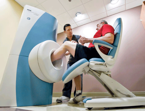
Revolutionizing Extremity Imaging: UFAI's Open MRI for the Foot and Ankle
Read More
-
 Listen Now
15 Summer Foot Care Tips to Put Your Best Feet Forward
Read More
-
 Listen Now
The Link Between Foot Health and Posture
Read More
-
 Listen Now
Why Are My Feet Different Sizes? It's More Common Than You Think
Read More
-
 Listen Now
Custom Orthotics vs. Over-the-Counter Inserts: Which Are Best for Your Feet?
Read More
-
 Listen Now
9 Running Tips from Sports Medicine Experts
Read More
-
 Listen Now
An Inside Look at Shockwave Therapy for Heel Pain, now available in Valencia, CA
Read More
-
 State-of-the-Art CT Scanning, Now in Our Office
Read More
-
 Listen Now
If the Shoe Fits, Wear it… Especially for Kids Shoes!
Read More
-
 Listen Now
Is Barefoot Running Better? Or are you Running Toward Injury?
Read More
-
 StimRouter: A Revolutionary Approach to Targeted Pain Relief
Read More


