- Home
- Foot & Ankle Conditions
- Bunions
Bunions: causes and advanced treatment options
What is a Bunion?
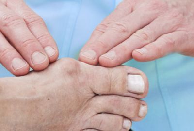
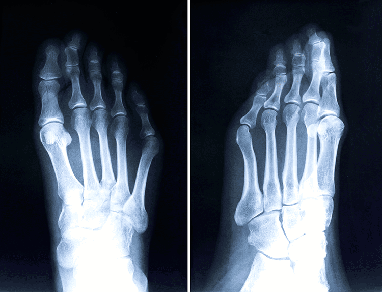
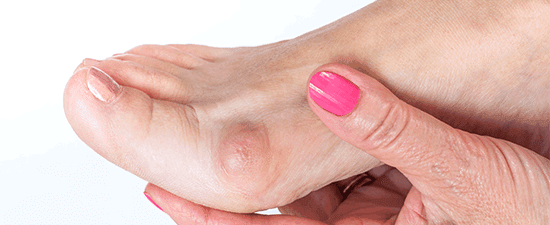
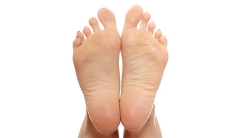
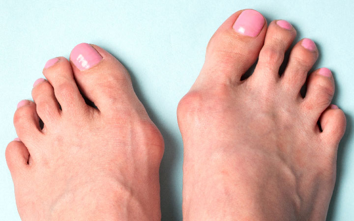
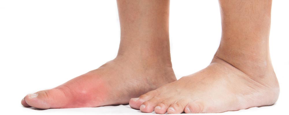
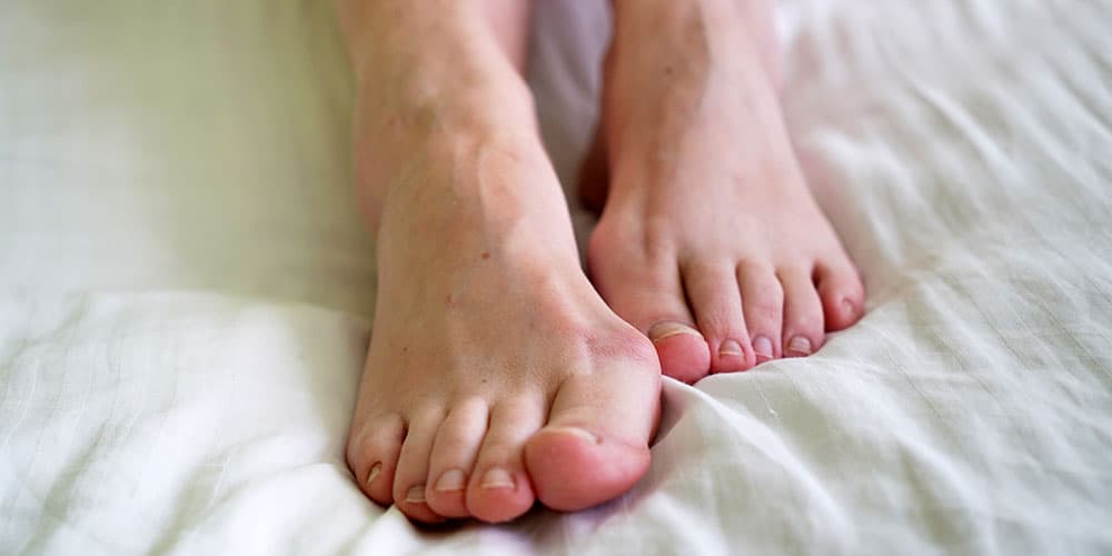
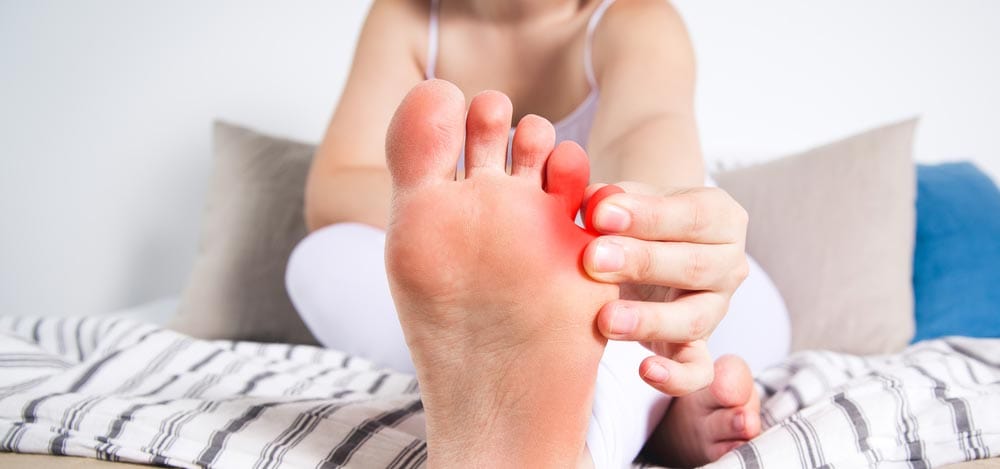
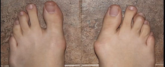
A bunion typically appears as a bony bump on the inside edge of the foot, where the innermost long bone of the foot (called the first metatarsal) meets the base of the big toe.
The visible bump appears when the first metatarsal tilts outward, causing the head of the bone to protrude. A bunion can also form at the base of the little toe, at which point it is known as a tailor's bunion.
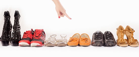
Causes of a bunion can include genetics, structural abnormalities of the foot, pregnancy, rheumatoid arthritis, and long-term wearing of high heels or pointy shoes.
- What sets UFAI's experts apart from other bunion surgeons?
- Our decades of surgical advances include:
- Our Bunion Surgeries are essentially painless and virtually scarless
- What happens if I do nothing about my bunion?
- Our state-of-the-art bunion correction procedures include:
- Bunion Revision Surgery
- Are there Non-Surgical Bunion Treatments?
- UFAI, the right choice for your bunion treatment
- Bunion FAQs
- Do bunion correctors work?
- Are bunions hereditary?
- Can bunions be corrected without surgery?
- I s bunion surgery covered by insurance?
- How long is recovery from bunion surgery?
- How do you relieve bunion pain when walking?
- Is bunion surgery painful?
- Can you get a bunion on your pinky toe?
- Can bunions cause leg pain?
- What are the best shoes for bunions?
- Is bunion surgery outpatient?

What sets UFAI's experts apart from other bunion surgeons?
Patients travel from all over the United States seeking University Foot & Ankle Institute’s advanced surgical techniques and attention to post-operative care that result in permanent correction and faster recovery with minimal pain and scarring.
Our decades of surgical advances include:
Dr. Barvarian discusses the Lapidus Forever Bunionectomy™️
- Development of the most advanced bunion surgery protocols, including minimally invasive Bunionplasty, Lapiplasty, and all-natural bunion correction.
- Pioneers of virtually scar-free surgical techniques.
- Over a decade of work in developing regenerative medicine protocols for foot and ankle surgery including Amniotic stem cells, Platelet Rich Plasma (PRP), Amniotic fluid allograft, and the new SoftWave Extracorporeal Shock Wave Therapy.
- Years of experience running and participating in research, development, and clinical trials of the latest bunion surgery hardware and technologies, including the Ossiofiber non-metal fixation material technology.
- Individualizing every bunion procedure to suit each patient’s anatomy and lifestyle (a critically important component when it comes to recovery). Our surgeons utilize special techniques for handling soft tissue and closing incisions so as to achieve a cosmetically pleasing outcome. We never take a “one-size-fits-all” approach to treating our patients.
- Teaching other physicians both in the United States and in Europe how to perform the refined surgical techniques that we’ve developed, achieving over 25,000 successful surgeries to date.
- Offering the most advanced technology to give our patients the best possible results.
Watch Our Videos About Bunions (12)
 01:58Bunion
 01:26Diane, Bunion Revision Surgery Patient
 01:17Beverly: Bunionectomy, Ankle Ligament & Tendon Repair, Patient Testimonial
 01:30Rene, Bunion Patient
 01:27Brenda, Bunion Patient
 01:41Kathy: Bunion, Hammertoe & Plantar Plate Repair, Patient Testimonial
 01:01Barbara, Bunion Surgery Patient
 01:20Bunion Revision Surgery Patient Testimonial, Carmella
 00:48What's a Bunion? The Doctor Explains
 01:41Kathy: Bunion, Hammertoe & Plantar Plate Repair Surgery
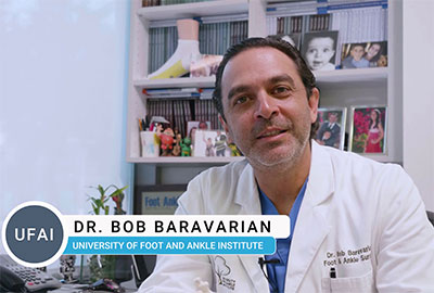 02:37The Forever Bunionectomy
 01:21Barbara, Bunion Patient Testimonial


Our Bunion Surgeries are essentially painless and virtually scarless
Many bunion sufferers avoid surgery because they fear a long and painful recovery and unsightly scarring. The state-of-the-art bunion correction protocols we helped develop result in an essentially painless recovery and inviable incisions. In fact, most patients tell us they wish they hadn’t waited so long to have their surgery.
Our surgeons are able to remove the pain and scarring from most surgeries by:
-
Taking extraordinary care in the handling of the soft tissue. It takes years of training and expertise to perform these types of dissections.
-
Using advanced fixation systems so the bones won’t have any micro-movement. This results in much less swelling and pain than with conventional fixation methods.
-
Utilizing long-acting local anesthesia with a proprietary “cocktail” of medications to control post-operative swelling and pain.
-
Incorporating regenerative medicine therapies into our treatment plans greatly stimulates healing and reduces recovery time.
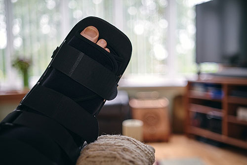
- Prescribing a special post-op boot which provides more stability and lessens the joint movement, resulting in less pain.
- Using post-op protocols to mitigate inflammation and pain without the need for excessive narcotic use.
The overwhelming majority of our patients rarely take more than one or two prescribed pain pills following surgery and can manage their post-op pain with over-the-counter anti-inflammatory medications and Tylenol.
What happens if I do nothing about my bunion?
Early-stage bunion deformities will almost always get worse over time. Complications from an untreated bunion include:
- Increased discomfort and pain. Bunion progression cannot be stopped. Your bunion will cause increased swelling and pain and the bump will appear more pronounced.
- Osteoarthritis or bursitis. As the joint shifts more and more, your joints are impacted and will eventually become very damaged. The cartilage will continue to deteriorate and a minimally invasive surgical procedure may no longer be an option for you.
- Crossover Toe. As more time passes, many patients see that their second toe begins to literally cross over the first toe. This causes pain, callusing, and even ulcers in your second toe. More invasive surgical procedures are now your only option.
Our state-of-the-art bunion correction procedures include:
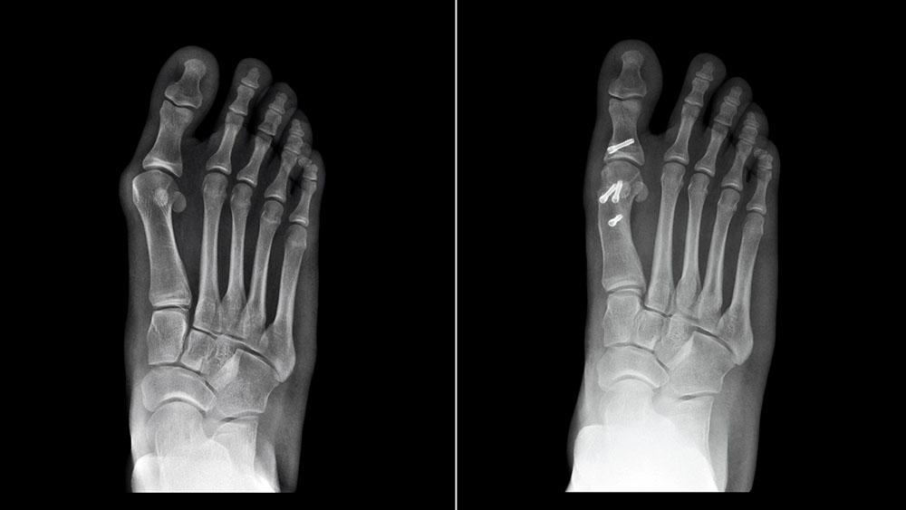
Bunionplasty minimally Invasive Bunion Surgery

Minimally Invasive Bunionectomy, after picture was taken six weeks post-surgery.
Our Bunionplasty technique combines the best parts of open bunionectomy (stabilizing the bone with hardware) with the best aspects of traditional minimally invasive bunionectomy (small incision surgery) and new technologies. By marrying the best of both approaches with new technologies, our Bunionsplasty is the ideal procedure for achieving the best potential outcomes.
Whereas traditional minimally invasive procedures utilize a thin, fragile pin to fixate cut bone, our Bunionplasty approach uses custom-designed fixation systems to provide a stronger more stable fixation that can be placed through a very small incision. One other unique feature of the Bunionplasty minimally invasive technique is that the bone can be shifted as far as necessary allowing us to correct any size.
Our type of fixation controls abnormal stress through the bone and holds both ends in the correct position until the bone fully heals. This results in excellent healing and limits non-union risks associated with traditional minimally invasive bunion surgeries.
Advantages of our Minimally Invasive Surgery (MIS) for bunions:
-
- Uses a tiny 1 cm incision
- Allows immediate weight-bearing after surgery
- Leaves a virtually undetectable scar
- Minimal swelling
- Minimal pain
- Often no need for physical therapy
- It is paired with Amniotic fluid allograft to speed up recovery and prevent internal scar tissue formation
- This surgery avoids future joint problems because it does not involve opening the joint
Osteotomy Bunionectomy

For moderate to severe bunions without joint instability.
Osteotomy procedures are one of the most commonly performed bunion surgeries. An osteotomy, or bone cut, is made at the tip of the first metatarsal. This “V” shaped bone cut allows the first metatarsal to shift so that it ends up parallel to the second metatarsal.
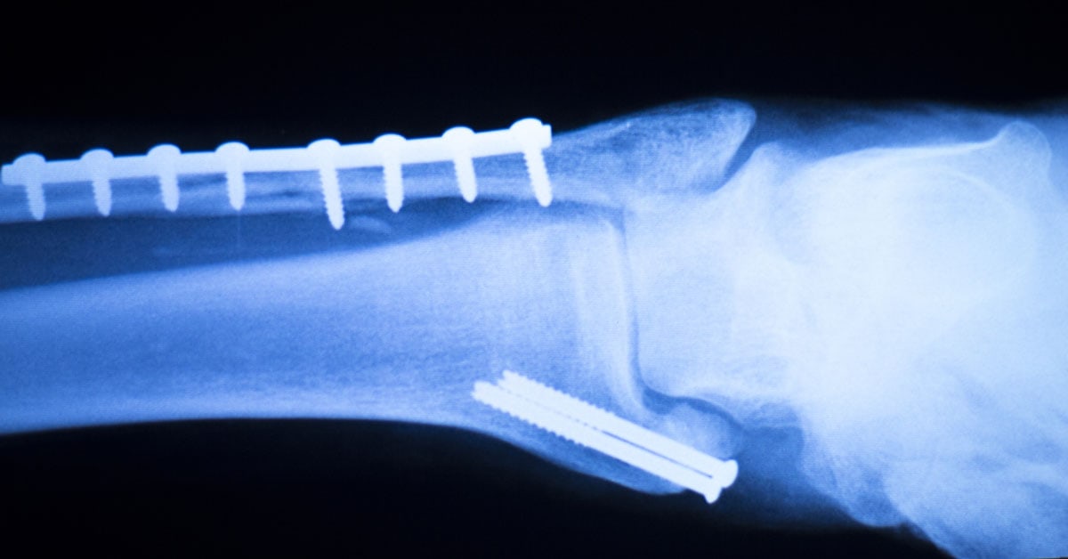
The metatarsal bones are held together in their newly corrected position with two screws. Although the screws may be redundant after the bone is healed, they are often left in place to avoid a re-occurrence of the bunion.
We are proud to offer a revolutionary non-metal fixation option for bunion surgery called OSSIOfiber®. OSSIOfiber is a bio-degradable polymer mixed with a mineral fiber that slowly grows into the human bone, reinforcing the bone healing site. It is eventually eliminated completely and replaced by the body’s own bone through natural cellular turnover. So, the material not only integrates but actually adds strength to the surgical site.
The osteotomy bunionectomy is a strong and predictable procedure and is commonly used on active patients younger than age 50.
Our surgeons revolutionized the traditional osteotomy procedure. The advantages of our Osteotomy Bunion Surgery include:
-
- Very small 3 cm incision
- Immediate weight-bearing
- A hidden incision along the side of the foot
- Plastic surgery closure techniques result in minimal scarring
- Early range of motion in recovery
- Cleans up spurs, if present
- Non-metal fixation option is available
-
Amniotic Fluid Allograft and PRP can be used to speed up recovery and reduce internal scar tissue formation and stiffness
Lapiplasty Bunion Correction

.
For moderate to severe bunions and the best option for those with joint laxity
Lapiplasty bunion correction is considered the Gold Standard for bunion surgery. For the right patient, this procedure has the highest success rates with essentially a 0% chance of the bunion returning.
This procedure has the lowest risk of re-occurrence of the bunion because the Lapiplasty addresses the source of the bunion deformity: a lax/hypermobile joint between the first metatarsal and the cuneiform bone located at the base of the big toe joint.
Lapiplasty surgery, considered to be the next-generation Lapidus Bunionectomy, uses patented instrumentation to manipulate the deviated bone back to its original position without cutting the bone, naturally realigning the foot and straightening the big toe, and alleviating the bump and accompanying pain.
Once proper alignment is achieved, Lapiplasty uses innovative bone fixation technology to secure the bones.
Advantages of our Lapiplasty Bunion Correction include:
-
- Permanent deformity correction with virtually no chance of reoccurrence
- Weight-bearing in just two weeks (versus 6-8 weeks with traditional approach)
- Plastic surgery closure techniques result in reduced scarring
- DynaForce Active Stabilization System foregoes the need for screws
- On-site CT scanner allows our doctors to detect the completion of bone fusion earlier, which gets our patients moving faster
- Our surgeons work directly with our physical therapists to offer our advanced PT protocol
Read more about Lapiplasty bunion correction here.
Bunionectomy Yelp Patient Review
The following five-star Yelp review was posted by University Foot and Ankle Bunion patient, Shabnam.

...From start to finish, before and after my bunion surgery, [my Dr.] and his staff took great care of me.
The surgery was fast and precise, with clean incisions and nearly painless recovery. I did not need to take any of my pain medication.
Everyone in his office is warm, friendly, and knowledgeable, which makes the entire experience very smooth and pleasant.
Click here to see Shabnam's full review on Yelp.com.
Bunion Revision Surgery
Many of our patients come to us after previous bunion surgeries at other facilities have proved unsuccessful. In fact, revisions account for almost one-third of our bunion surgeries. It pains us that so many people need to go through surgery a second time.
Failed bunion surgeries are discovered when new complications appear, including recurrence of the bunion, an excessively short big toe, stiffness in the toe joint, arthritis,
It is essential to choose your revision surgeon wisely. Bunion revision surgery requires the necessary experience and skills to understand and address the shortcomings of the original surgery.
Are there Non-Surgical Bunion Treatments?
While you technically can’t fix a bunion without surgery, there are ways to slow or even stop the progression of the deformity. Custom orthotics, shoe modifications, exercise, stretching, bunion pads, and splints are commonly prescribed conservative treatments.
Learn more about our non-surgical bunion treatments here.
While You're Here...
UFAI, the right choice for your bunion treatment
As an internationally recognized Center of Excellence known for our expertise in bunion surgery techniques and research, our surgical team is able to offer a treatment plan that will address the deformity, correct the underlying cause of the bunion, and prevent a recurrence.
Our orthopedic surgeons further advance bunion surgery by adding stem cells, amniotic fluid allograft, and PRP. These regenerative medicines may be added during surgery to promote bone healing, speed up recovery, and reduce internal scar tissue and stiffness.
With over 25,000 successful procedures performed to date, our success speaks for itself.
For a consultation, please call (877) 736-6001 or make an appointment online now.
University Foot and Ankle Institute is conveniently located throughout Southern California and the Los Angeles area. Our foot and ankle surgeons are available at locations in or near Santa Monica, Beverly Hills, West Los Angeles, Sherman Oaks, the San Fernando Valley, El Segundo, the South Bay, LAX, Calabasas, Agoura Hills, Westlake Village, Valencia, Santa Clarita, and Santa Barbara.
Bunion FAQs
Do bunion correctors work?
While widely available and marketed as a solution for bunions, bunion correctors often have limited effectiveness. It's essential to understand that bunions are a structural issue, and while these devices may provide some temporary relief, they are unlikely to correct the root cause of the deformity.
Are bunions hereditary?
Yes, genetics plays a significant role in the development of bunions. If your parents or grandparents had bunions, you may have an increased risk of developing them as well. However, it's important to note that other factors like footwear, foot mechanics, and lifestyle can also contribute to their development.
Can bunions be corrected without surgery?
Bunions can often be managed and improved without surgery, especially if they are detected early. However, the effectiveness of these approaches varies depending on the severity of the bunion and individual factors.
Here are some non-surgical methods to consider:
- Footwear
- Orthotics
- Padding and taping
- Physical therapy and exercises
- Medications
Is bunion surgery covered by insurance?
In many cases, bunion surgery is covered by insurance when it is deemed medically necessary. Medical necessity is typically based on the severity of the bunion, the impact it has on your daily life, and the failure of conservative treatments to provide relief.
Keep in mind that cosmetic bunion surgery, which is performed solely for aesthetic reasons, is typically not covered by insurance.
How long is recovery from bunion surgery?
The recovery time for bunion surgery may take from six months to a year or longer to achieve full recovery depending on the extent of the surgery, individual healing factors, overall health, and how well post-operative care instructions are followed.
How do you relieve bunion pain when walking?
There are several strategies to help relieve foot pain when walking:
- Proper footwear: Wear shoes with a wide toe box and good arch. Avoid high-heeled shoes or shoes with narrow or pointed toes.
- Shoe modifications: Consider getting custom-made orthotic insoles prescribed by a podiatrist or over-the-counter arch support inserts. Use bunion pads or cushions to provide a protective barrier between the bunion and your footwear. These can help reduce friction and pressure.
- Nonsteroidal Anti-Inflammatory Drugs (NSAIDs)
- Ice therapy: Applying ice to the bunion for 15-20 minutes at a time can help reduce swelling and alleviate pain.
- Rest and elevation: Give your feet adequate rest, especially if the bunion pain is severe. Elevating your foot when resting can help reduce swelling.
- Physical therapy
Is bunion surgery painful?
Despite pre-surgical fear of pain and recovery, virtually all our patients report they essentially no pain and wish that they had surgical correction sooner and hadn’t put it off as long as they did.
Our advances in surgical techniques drastically reduce recovery time, often allowing you to bear weight immediately.
Can you get a bunion on your pinky toe?
Bunions on the pinky toe are less common but not impossible. When a bony bump or prominence forms on the outer side of the foot near the base of the pinky toe, it is referred to as a tailor's bunion or bunionette.
Can bunions cause leg pain?
Bunions themselves are primarily localized to the foot and affect the joint at the base of the big toe. However, in some cases, bunions can indirectly contribute to leg pain or discomfort due to altered gait and joint stress.
What are the best shoes for bunions?
Choosing the right shoes for bunions is crucial to minimize discomfort and prevent further irritation. Here are some features to look for when selecting the best shoes for bunions:
- Wide Toe Box
- Low Heel or No Heel
- Flexible and Cushioned Soles
- Arch Support
- Adjustable Straps or Laces
- Soft and Breathable Materials
- Removable Insoles
- Orthopedic or Bunion-Specific Shoes
Is bunion surgery outpatient?
Yes, typically, bunion surgery is performed on an outpatient basis.
-
Foot and Ankle Surgeon and Director of University Foot and Ankle Institute
Dr. Bob Baravaria DPM, FACFAS is a Board-Certified Podiatric Foot and Ankle Specialist. He is an assistant clinical professor at the UCLA School of Medicine and serves as Director of University Foot and Ankle Institute.
Dr. Baravarian has been involved in athletics his entire life and played competitive tennis in high school and college. He has an interest in sports medicine, arthritis therapy, and trauma/reconstructive surgery of the foot and ankle. He is also fluent in five languages (English, French, Spanish, Farsi, and Hebrew),

Read Our Blog Articles About Bunions
- The Link Between Foot Health and Posture
- Understanding Tailor's Bunions: Causes, Symptoms, and Solutions
- Bunion surgery gone wrong: what happens when your bunion surgery fails?
- 12 Tips to Prepare Your Home for Bunion Surgery Recovery
- Do Bunion Surgery Techniques Outlive their Usefulness? Yes!
- Non-Surgical Bunion Treatments
- Bunions vs. Big Toe Arthritis, What's the Difference?
- Little Toe Hurts? Four Things to Know About Pinky Toe Pain
- Got Big Toe Bumps and Lumps? Here’s 5 Things You Need to Know
- 16 Myths About Bunion Surgery Debunked!
- Metal Surgical Screws and Pins May Become Thing of the Past
- Finding the Best Bunion Surgeon: What You Need to Know
- 11 Common Foot Lumps and Bumps and What To Do About Them
- How to Choose Good Bunion Shoes (Even High Heels)!
- Beat the 6 Most Common Walking Pains
- The Least Instagram-able Baby Bump Ever: Pregnancy Bunions
 Dr. Gary Brisken has been my doctor going on two years. He's a very kind and thoughtful physician. The heath of my feet has imp...Mark P.
 Great experience. Great communication. Great direction for my care. Very happy I chose to go with this particular doctor and o...Christopher R.
- Great service and care. Highly recommend Dr. Franson.David B.
- If you have to go see a Doctor than this is a great experience.Frank M.
- My doctor was great. Really greatRudolph B.
- Good.David E.
 Dr Franson is amazing…. He’s done 3 surgical procedures on me, helped me with bilateral plantar fasciitis, a broken foot and a ...Rose R.
- Your Santa Barbara office and Dr. Johnson always give me excellent care!Jayne A.
- My experience at UFAI was , has and still is a perfect example of how all medical visits should be . No not just medical but an...Reznick L.
- Dr. Gina Nalbadian was amazing!! I came in with an emergency foot situation and she had wonderful bedside manner and resolved m...Danielle C.
- I was frustrated that after 3 weeks I still hadn’t heard back about my PT referral status. And I did sit in a room for over 30 ...Sarah C.
- I’m very pleased with Dr. Kelman.Alan S.
-
 Understanding Tailor's Bunions: Causes, Symptoms, and Solutions
Read More
Understanding Tailor's Bunions: Causes, Symptoms, and Solutions
Read More
-
 Listen Now
Beat the 6 Most Common Walking Pains
Read More
Listen Now
Beat the 6 Most Common Walking Pains
Read More
-
 Listen Now
Finding the Best Bunion Surgeon: What You Need to Know
Read More
Listen Now
Finding the Best Bunion Surgeon: What You Need to Know
Read More
-
 Listen Now
11 Common Foot Lumps and Bumps and What To Do About Them
Read More
Listen Now
11 Common Foot Lumps and Bumps and What To Do About Them
Read More
-
 Listen Now
12 Tips to Prepare Your Home for Bunion Surgery Recovery
Read More
Listen Now
12 Tips to Prepare Your Home for Bunion Surgery Recovery
Read More
-
 Listen Now
Bunions vs. Big Toe Arthritis, What's the Difference?
Read More
Listen Now
Bunions vs. Big Toe Arthritis, What's the Difference?
Read More
-
 Listen Now
Non-Surgical Bunion Treatments
Read More
Listen Now
Non-Surgical Bunion Treatments
Read More
-
 Listen Now
The Link Between Foot Health and Posture
Read More
Listen Now
The Link Between Foot Health and Posture
Read More
-
 Listen Now
Got Big Toe Bumps and Lumps? Here’s 5 Things You Need to Know
Read More
Listen Now
Got Big Toe Bumps and Lumps? Here’s 5 Things You Need to Know
Read More
-
 Listen Now
Bunion surgery gone wrong: what happens when your bunion surgery fails?
Read More
Listen Now
Bunion surgery gone wrong: what happens when your bunion surgery fails?
Read More
-
 Listen Now
Do Bunion Surgery Techniques Outlive their Usefulness? Yes!
Read More
Listen Now
Do Bunion Surgery Techniques Outlive their Usefulness? Yes!
Read More
-
 Listen Now
Metal Surgical Screws and Pins May Become Thing of the Past
Read More
Listen Now
Metal Surgical Screws and Pins May Become Thing of the Past
Read More
-
 Listen Now
Little Toe Hurts? Four Things to Know About Pinky Toe Pain
Read More
Listen Now
Little Toe Hurts? Four Things to Know About Pinky Toe Pain
Read More
-
 Listen Now
How to Choose Good Bunion Shoes (Even High Heels)!
Read More
Listen Now
How to Choose Good Bunion Shoes (Even High Heels)!
Read More
-
 Listen Now
16 Myths About Bunion Surgery Debunked!
Read More
Listen Now
16 Myths About Bunion Surgery Debunked!
Read More


