Accessory Navicular: causes, symptoms and treatment

Updated 12/28/2023
Accessory Navicular, University Foot and Ankle Institute

What is an accessory navicular? 

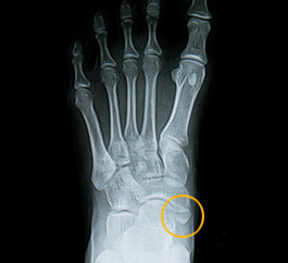
The accessory navicular (os tibiale externum) is an extra piece of cartilage or bone located midfoot just above the arch on the inside of the foot.


This ossicle (extra bone) is usually encased in the posterior tibial tendon, which attaches the foot's inside (medial) arch to the calf muscle. Accessory navicular is commonly seen in pediatric patients.


Most people do not have an accessory navicular. People who do have one are born with this condition and may go their whole lives without realizing it. Others will experience pain and foot deformities.

Why does an accessory navicular require bone treatment? 

An accessory navicular may not cause problems in every patient. However, trauma (such as a foot or ankle sprain) can sometimes cause the accessory navicular to pull apart from the main bone. When two bones rub together, they can become inflamed.


Once the tiny bone is inflamed, it is difficult to reduce the irritation without medical intervention.


An accessory navicular can also be the cause of flatfeet. The small extra bone can put strain on or weaken the posterior tibial tendon. Without the support of the tendon, the arch of the foot collapses.


Accessory navicular syndrome can also be caused by ill-fitting shoes and overuse.


What are the symptoms of accessory navicular syndrome? 

Most people with accessory navicular syndrome start noticing symptoms in their teens when their youthful cartilage develops into mature bone tissue. Signs of accessory navicular syndrome include:


  • A bony protrusion on the inside midfoot, just above the arch.
  • Swelling and redness around the protrusion.
  • Persistent or throbbing pain in the arch, especially during or after exercise.


How is accessory navicular syndrome diagnosed?

The first step in diagnosing any foot pain is a physical exam with one of our foot and ankle surgeons. 


Our doctors will examine the shape of the foot, observe the foot’s mechanics while walking, and check for any abnormal bony prominences attached to the tuberosity of the navicular. X-rays and MRIs allow us to see the foot bones and surrounding soft tissue. 


A physical exam accompanied by imaging allows us to rule out other potential causes of foot pain such as plantar fasciitis or tibialis posterior tendonitis.


The three types of accessory navicular 

During diagnosis, we will also determine what type of accessory navicular you have, which will also inform your treatment options.


  • Type 1: A type 1 accessory navicular is small and either round or oval and sits within the posterior tibial tendon. These are also called sesamoids.
  • Type 2: A type 2 accessory navicular is bigger than type 1, bullet-shaped, and attached to the tuberosity of the navicular by a synchondrosis (a cartilaginous joint). This is the type most likely to have symptoms.
  • Type 3: A type 3 accessory navicular is quite prominent and is described as horn shaped. A type 3 accessory navicular is believed to be a type 2 that has fused.


What are accessory navicular treatment options? 

Some cases of accessory navicular syndrome can be healed conservatively with nonsurgical treatment. At University Foot & Ankle Institute, our podiatrists opt for conservative treatment whenever possible.


Conservative treatment methods may include:


Icing 

Wrap a small bag of ice in a paper towel or plastic bag and apply it to the area for 15-20 minutes as needed to decrease pain and swelling.


Medication 

Our doctors recommend using nonsteroidal anti-inflammatory drugs (NSAIDs) to manage the pain and decrease inflammation. You can choose from several over-the-counter options that contain ibuprofen, such as Advil or Motrin.


Immobilization 

Our specialists may set your foot in a cast or removable walking boot for two to three weeks to give the inflammation time to heal. Oral or injected steroid medications may be used in combination with immobilization to reduce pain and inflammation.


Physical therapy 

Our podiatrists can show you how to perform daily physical therapy exercises to strengthen the muscles, reduce pain, and avoid future problems.


Orthotics 

Our foot experts can fit you for custom orthotic inserts to provide increased arch support, stabilize ligaments with insoles, and reduce pain during physical activity.


What is accessory navicular bone surgery? 

The University Foot and Ankle Institute specialists may recommend surgical intervention if conservative treatments cannot manage symptoms. The extra bone does nothing to help the foot’s mechanics, so surgical removal could effectively relieve pain and restore proper foot function.


Known as the Kidner procedure, this surgical excision technique involves removing the accessory navicular bone and repositioning the posterior tibial tendon. The Kidner procedure is performed under general or spinal anesthesia. Our surgeon makes an incision along the inner side of the foot just above the arch, which exposes the accessory bone and the posterior tibial tendon. Our doctors remove the accessory navicular and reattach the tendon to the prominent navicular bone.


The Kidner procedure is a relatively low-risk surgery with a high chance of normal functioning after recovery. The patient can expect to be resting in bed in a surgical cast for about two to three weeks and then work their way up to weight-bearing in a boot for two to four additional weeks. After six weeks, most patients can bear total weight on the foot and begin physical therapy. They typically return to their normal foot function after six months of rehabilitation.


University Foot & Ankle Institute is ideal for accessory navicular treatment 

You don't have to live with foot pain. There is so much innovation in podiatric medicine today, and our board-certified orthopaedic surgeons are at the forefront. With decades of combined experience and the highest success rates in the nation, we have effectively treated over 200,000 patients.


Foot and ankle pain is not normal. And ignoring it will likely cause your condition to worsen and become harder to treat. 


The University Foot and Ankle Institute (UFAI) is well known as one of the nation's most technologically advanced orthopedic practices (including sports medicine). While other practices say they know about "the latest and greatest," UFAI has been researching, running orthopedic clinical trials, and performing these techniques for decades. 


We've been a leader in regenerative medicine for well over 15 years. This includes platelet-rich plasma therapy, stem cell therapy, and cartilage replacement. No other foot and ankle practice has more experience or success in developing and using human regenerative medicine.


University Foot and Ankle Institute is conveniently located throughout Southern California and the Los Angeles area. Our foot doctors are available at locations in or near Santa Monica, Beverly Hills, West Los Angeles, Manhattan Beach, Northridge, Downtown Los Angeles, Westlake Village, Santa Barbara, and Valencia.


References

Accessory Navicular Syndrome – Navicular Bone Pain | Foot Health Facts - Foot Health Facts

https://www.foothealthfacts.org/conditions/accessory-navicular-syndrome-(1)


Foot Health Facts | Foot and Ankle Surgeons - Podiatrists - Foot Health Facts

http://www.foothealthfacts.org


Accessory Navicular Syndrome – Navicular Bone Pain | Foot Health Facts - Foot Health Facts

http://www.foothealthfacts.org/footankleinfo/acc

19,483 Total 1st Party Reviews / 4.9 out of 5 Stars
  • Google
    Dr. Franson was very professional and friendly. I would definitely recommend him.
    Sherry G.
  • GatherUp
    Great experience. Great communication. Great direction for my care. Very happy I chose to go with this particular doctor and o...
    Christopher R.
  • GatherUp
    Great service and care. Highly recommend Dr. Franson.
    David B.
  • GatherUp
    If you have to go see a Doctor than this is a great experience.
    Frank M.
  • GatherUp
    My doctor was great. Really great
    Rudolph B.
  • Google
    I am a diabetic. I dropped a 2 drawer metal file cabinet on my big toe. Due to the diabetes I knew I had to have it checked o...
    Darcy T.
  • GatherUp
    Good.
    David E.
  • GatherUp
    Your Santa Barbara office and Dr. Johnson always give me excellent care!
    Jayne A.
  • GatherUp
    Dr. Gina Nalbadian was amazing!! I came in with an emergency foot situation and she had wonderful bedside manner and resolved m...
    Danielle C.
  • GatherUp
    I was frustrated that after 3 weeks I still hadn’t heard back about my PT referral status. And I did sit in a room for over 30 ...
    Sarah C.
  • GatherUp
    I’m very pleased with Dr. Kelman.
    Alan S.
  • Google
    Great care....Been going here for years!
    Mary B.
Same Day Appointments
Now Available!

Or call 877-989-9110

24 hours a day, 7 days a week