University Foot and Ankle Institute has its own foot and ankle MRI scanner conveniently located in the Santa Monica facility, offering our patients several substantial advantages.
Our ONI MSK Extreme MRI scanner was designed so our doctors can perform high-field (which basically means “extremely clear”) diagnostic images in an environment optimized for our patient’s comfort.
“Open MRI” units (where your body does not completely go into a very closed-in feeling tube) usually have inferior image quality compared to traditional MRI machines. However, UFAI’s ONI Open MRI is the world’s only open extremity MRI scanner to offer the quality of a traditional MRI, and because only your foot and ankle have to be inside the “imaging tube,” it is truly fantastic.
How does UFAI’s Foot and Ankle MRI differ from scans at other imaging centers?
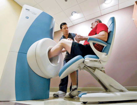
Our MRI scanner is specifically designed for imaging of the extremities, therefore only the joint being imaged is inside the scanner and the patient is comfortably seated outside the machine. This anatomy-specific MRI scanner provides a quiet, comfortable, non-claustrophobic experience for our patients. This state-of-the-art scanner is not only found at University Foot and Ankle Institute but at top hospitals, imaging centers, and the top academic hospitals in the world.
In addition to assessing the ankle and foot bones, the ONI provides our podiatrists with extremely accurate images of small structure conditions, such as ligaments and tendons.
University Foot and Ankle Institute podiatrists believe in diagnosing thorough scans and capture more images than similar facilities.
The advantages of the Open MRI study at University Foot and Ankle Institute
MRI’s at University Foot and Ankle are very different than those taken at other facilities. Insurance companies will only pay for a pre-set amount of time for your foot or ankle to be scanned. This amount of time is only enough to get the minimum number of views of your injury to make a standard diagnosis. It usually consists of just four views, which often is inadequate for the most accurate diagnosis.
There are three main advantages to having an MRI stud at University Foot and Ankle Institute’s Open Foot and Ankle MRI
- Our MRI is specifically made for the foot and ankle. Thus your whole body (especially your head) never goes go into any “tube”, only your foot does, so it’s really good in that way. Patients who have done it tell us, believe it or not, it’s actually pleasant.
- We take extra views of the foot with their own MRI machine when, as mentioned above, other facilities do not. Plus we don’t charge for those extra views because we can only charge a certain amount for an MRI anyway. But we get the extra views because it absolutely helps your doctor diagnostically, so it’s standard operating procedure at UFAI. Other facilities simply don’t do that.
- We can tell immediately if the MRI has any issues, such as the foot moved during the study so some views need to be redone. We check the images before we send you on your way so you never have to come back if there’s a problem. We just retake those views immediately. Not that there are common issues with other imaging center’s MRI’s, that’s not the case. But it has happened, and when it does, it’s obviously not fun to have to go back and have it redone.
Dr. Briskin is a Diplomat of the American Board of Podiatric Surgery and a Fellow of the American College of Foot and Ankle Surgeons. He also serves as an assistant clinical professor at the UCLA School of Medicine.
- Revolutionizing Extremity Imaging: UFAI’s Open MRI for the Foot and Ankle - October 21, 2023
- Youth Sports and Heel Pain: Should Kids Play with Pain? - April 4, 2023
- All About Foot Arch Pain and Foot Arch Cramps - March 15, 2021