- Home
- UFAI in the News
- UFAI Medical Journals
- Intermetatarsal Compression Neuritis
Intermetatarsal Compression Neuritis
- Published 7/3/2006
- Last Reviewed 7/3/2006

Written by Justin Franson, DPM and Bob Baravarian, DPM
Intermetatarsal neuroma has been well described in the literature over the years. Although the condition often bears the name of Morton, who described the condition in 1876 [1], the original description has been attributed to Durlacher in 1845 [2].
‘‘Morton’s neuroma’’ is not a true neuroma, although the use of the term has been perpetuated in the literature. A more accurate description of this condition is intermetatarsal compression neuritis (ICN), which will be used in this article. This forefoot pathology can be a disabling condition; individuals who don’t respond well to initial treatments are left with persistent pain and frustration. The treatment options have evolved over the years and there are now more options available to the practitioner than the simple conservative treatments and surgical resection. It is important to understand the condition well, and pursue new treatment protocols that can lead to improved outcomes.
Occurrence
Although the condition can be found in any individual, ICN is more common in middle-aged females, especially those who frequently wear high-heeled and pointed-toe shoes [3]. The abnormal biomechanical forces and toe crowding caused by many fashionable shoes have been suggested as etiological factors in the development of this condition.
It is generally accepted that the third intermetatarsal space is the anatomic location most susceptible to ICN [3]. Dockery [4] reported results on 100 neuromas, of which 81 cases involved the third intermetatarsal space. However, in one long-term follow-up study of ICN, 67% of 70 neuromas treated were located in the second intermetatarsal space [5]. First and fourth interspace neuromas are rare [3].
Anatomy
The intermetatarsal nerves that pass between the metatarsal heads are branches from the medial and lateral plantar nerves, the continuation of the tibial nerve. The third intermetatarsal space receives nerve contributions from a branch of the medial and lateral plantar nerves. It has been suggested that the nerve is larger in this area because of the two nerve branches that join here, which provides an anatomic explanation for the more common occurrence of neuroma in the third intermetatarsal space. Furthermore, there is a noted medial and lateral column to the foot where the third metatarsal is connected to a stable cuneiform; the fourth metatarsal is connected to a more mobile cubiod. This may facilitate greater motion at the third interspace level between the metatarsal heads, which results in nerve irritation and fibrosis.
As they near the distal level of the metatarsals, the common digital nerves pass beneath, or plantar to, the deep transverse intermetatarsal ligament. The nerves then branch into the proper plantar digital nerves, and supply sensory innervation to the toes.
Etiology
Although there has been some difference of opinon regarding the pathoetiology of this condition, it is frequently attributed to a mechanical entrapment neuropathy as the nerves pass between the metatarsal heads. It has been suggested that the histopathologic appearance of neuromas are related to two types of microtrauma: stretching and compression [6].
Intermetatarsal compression neuritis can occur in a pronated, supinated, or neutral foot. In the Cavus foot type, it has been suggested that there is increased tension on the plantar fascia and the deep transverse intermetatar- sal ligament [7]. This tension in the deep fascia structures render them less forgiving and place a more rigid structure under which the nerve will pass. When the toes are placed in a relative dorsiflexed position by high- heeled shoes, or as they dorsiflex in the propulsive phase of the gait cycle, the intermetatarsal nerves are placed against the tight, deep transverse metatarsal ligament. Routine radiographs obtained in the work-up of ICN in relation to cavus feet often shows a relative proximity of the metatarsal heads in the affected intermetatarsal space.
Pronated and neutral feet may also be affected with ICN. The authors have observed that in neutral or pronated feet, there is often hypermotion of the metatarsal heads (noted as ‘‘a loose bag of bones’’) that results in excess compression and motion at the metatarsal heads and neuritis or fibro- sis of the nerve.
Diagnosis
A careful review of the patient’s description of the symptoms coupled with a physical examination will usually be sufficient to arrive at the diagnosis of neuroma. Symptoms can include pain at the ball of the foot, burning pain, tingling, numbness in the toes, pain radiating into the toes or proximally into the arch, cramping, a shooting or stabbing pain, or a feeling like the patient is walking on a pebble or knot. Neuromas are not always easy to distinguish from other conditions that affect this anatomic area of the foot. Differential diagnosis can include tarsal tunnel syndrome or peripheral neuropathy, metatarsal stress fracture, tear of the plantar plate, plantar metatarsophalangeal joint capsulitis, avascular necrosis of the meta- tarsal head, metatarsal bone tumor, soft tissue mass, bursitis, flexor digito- rum tendonitis, and hammertoes that cause a retrograde buckling force and induce metatarsalgia.
Physical examination includes direct, deep palpation of the intermetatarsal space. Pain can usually be induced with deep intermetatarsal palpation, and simultaneous side-to-side compression of the metatarsals. This will often produce the ‘‘Mulder’s sign,’’ a palpable and sometimes audible click of the neuroma between the metatarsal heads. Diagnostic injections at the distal intermetatarsal space using a local anesthetic can help differentiate the distal ICN from a more proximal cause, such as tarsal tunnel syndrome or peripheral neuropathy.
Pressure specified sensory testing (PSSD) is a newer diagnostic tool which can be used to help rule out the presence of tarsal tunnel or other peripheral compression neuropathy. It can be used to evaluate and confirm an ICN, however, it is more difficult to examine the lesser digital nerve function. A more complete discussion of PSSD testing is given in the neurosensory testing article by Soomekh and colleagues elsewhere in this issue.
Imaging
Evaluation of ICN will often lead to routine radiographs, which can help rule-out other pathology. It is common to see a relative closer proximity of the adjacent metatarsal heads in the affected intermetatarsal space.
Although the diagnosis of ICN is largely based on clinical suspicion after a review of the patient’s symptomatology and a thorough clinical examina- tion, imaging is sometimes used pre-operatively. There can, however, be false negatives and false positives that may confuse the decision-making progress. The use of computerized tomography, diagnostic ultrasound, and MRI have been presented in the literature [8–10] as acceptable forms of imaging that can yield relatively accurate images of the affected nerve, although in most cases, they are used to rule out other possible causes of pain and symptoms. To complicate diagnosis, ICN does not always produce the bulbous swelling and nerve enlargement sometimes seen with this condition.
Ultrasound can be a very reliable modality to detect the pathology. This does rely, however, on an experienced ultrasonographer as well as a high-quality device. Ultrasound is less expensive than MRI or CT, and does not expose the patient to radiation. The procedure is performed by placing the probe parallel and just distal to the metatarsal heads. The potential involved interspace is then pressed from a dorsal motion and an enlarged nerve branch may be noted plantar to the transverse intermetatarsal liga- ment. A second position is to place the probe in a linear fashion, dorsally between the metatarsal heads, and observe the nerve in a longitudinal fash- ion. This is far more difficult to perform and requires a highly skilled ultrasonographer.
Multiple papers have discussed ultrasound diagnosis of ICN in the foot. A paper by Mendicino and colleagues [9] reported accurate detection of the presence, location, and size of the neuroma, as high as 95%–98%. However, in another study that compared pre-operative imaging with surgical find- ings, approximately half of the neuromas were missed by ultrasound and MRI [11].
Magnetic resonance imaging can produce the finest soft-tissue contrast. The edematous nerve is best seen on a T1-weighted image, and will be visualized as a localized mass with lower signal intensity. However, the edema- tous region can also be missed if the slices of imaging are not close enough together and the region is not filmed properly.
Pathology
Differing information exists in the literature regarding the pathologic findings in the presence of symptomatic neuromas.
In a 2000 study by Morscher and colleagues [12], the morphology and histological findings of ICN were checked. The excised neuromas were compared with biopsies of similar cadaver intermetatarsal nerves. They found no qualitative differences in the histomorphological examination of neuromas as compared with the cadaver specimens. The study did observe differences in the nerve diameter, although increased diameter is not always seen. This study further showed that the 17 smallest diameter nerves they exam- ined were from autopsy specimens, and the 5 largest diameter nerves were from resected neuromas. The medium diameter nerves showed a wide over-lap of the neuroma and the normal cadaver specimens. Based on this information, they concluded that MRI and ultrasonography are not necessary when deciding about surgical intervention, and were less reliable than diagnostic local anesthetic injections [12].
The only problem with local injections, is differentiation of plantar plate tears from neuroma pain, because both will improve with injection. However, with proper clinical correlation, a differentiation may be made.
Conservative treatment
The conservative approach to the treatment of neuromas includes shoe modifications, padding, orthotics, cortisone injections, and serial alcohol sclerosing injections [8]. The response to the conservative treatment of ICN is often directly related to the persistence, focused treatment, and creativity of physician. Those who more aggressively use the conservative measures are more likely to alleviate pain without surgical intervention. An article in Foot and Ankle International [6] concluded that orthotics that control rearfoot pronation produced no significant benefit in treating this condition.
It is well known that ICN is more commonly found in females, often middle-aged women. Shoes have been implicated as a causative factor in the development of neuromas. Shoe modifications should be strongly encouraged: wide toe box shoes and flat, heel-less shoes are best.
Cortisone injections are a well-accepted conservative treatment of the symptoms of neuroma, although they will likely not have a lasting effect unless other modifications are instituted. The injected steroid provides an anti- inflammatory mechanism at the area of pathology.
A series of dilute alcohol slerosing injections has been proposed as a favorable conservative treatment. The injected solution, a dehydrated alcohol mixed with local anesthetic with epinephrine, has a high affinity for nerve tissue, and produces a chemical neurolysis. Dockery’s [8] paper reported that 89% of patients improved. Long-term outcomes with this treatment have yet to be presented. The authors have been unable to reproduce such a high rate of success using the alcohol sclerosing injections. In spite of this, it is a useful option, especially for patients who are not surgical candi- dates, or prefer to avoid surgical intervention.
Custom orthotics has been a standard treatment option for intermetatar- sal neuritis. Although the effectiveness of this treatment has been questioned in the literature [6], the authors’ clinical experience has been that orthotics can be helpful. By controlling the rearfoot and midtarsal joints, abnormal forefoot compensation can be alleviated. Placement of padding can also function to spread the metatarsals and cushion the ball of the foot.
Surgical treatment
When a person has failed conservative treatment for a forefoot neuroma, surgery can be considered to alleviate pain and treat the condition. Several different surgical approaches have been described in the literature.
Neurectomy
The traditional surgical approach has been to perform a surgical excision of the affected nerve proximal to the metatarsal heads. This can be accomplished using multiple incisional approaches: dorsal longitudinal, plantar transverse, and plantar longitudinal. The excision of the nerve has produced varied results. One study observed a 93% patient satisfaction rate over the long-term (average 4.8 years), after dorsal longitudinal incision was used for neuroma excision [5]. The downside to performing nerve excision is the post- operative dysesthesias and the potential for a painful stump neuroma.
Carbon dioxide laser
A study by Wasserman [13] discussed the use and clinical outcome of a carbon dioxide laser in neuroma therapy. The author relates excellent re- sults; of 50 randomly sampled cases, there was only a single complication, and that was related to use of the ankle tourniquet. One noted benefit of the laser was that the nerve stump was sealed. Their patients were able to return to normal ambulation faster than with the conventional surgical approach.
Neurolysis
Another surgical approach is to perform a neurolysis, or surgical nerve decompression. This can be done endoscopically or through an open approach. The authors’ preference is through an open approach, so they can directly see the area. This also facilitates opening the endoneurium. The procedure is performed through a 2–3 cm dorsally based incision, placed in the noted interspace. Blunt dissection is carried to the level of the intermetatarsal ligament and a freer elevator or straight hemo- stat is placed plantar to the ligament. The ligament is then linearly incised and cauterized at the edges with a bipolar style cautery to prevent damage to the nerve. Any scar and fibrosis about the nerve is then released under high-power magnification. Suturing the skin is performed with the surgeon’s choice of closure. On the day of surgery the patient is allowed to apply weight in a surgical shoe and can move into a tennis shoe at 1 week when wound healing has progressed. Physical therapy is started at 2 weeks to break up fibrosis.
A recent article discussed intermetatarsal nerve decompression with reloca- tion dorsal to the intermetatarsal ligament of the digital nerve [14]. The article presented the surgical outcomes of 82 feet, 95% of which had complete reso- lution of neuritic symptoms within an average of 7 days following surgery.
Review of neurolysis results
The Foot and Ankle Institute Network has followed the progress of the authors’ first 30 cases of intermetatarsal nerve decompression over the past year. The procedure is performed through a linear interspace incision for each involved interspace. The incision is deepened and the intermetatarsal ligament is released. Careful dissection of the nerve is then performed; any fibrosis about the nerve is debrided, and the nerve is decompressed as needed under visualization with high-power magnification loupes.
The overall findings have been excellent: pain relief resulted in all cases. Three 3 of the 30 cases have had some level of continued pain after neurolysis. One case had previous alcohol injections for three sessions before neurolysis; a neurectomy was eventually performed because the nerve pain did not resolve completely following decompression. A second case had no pain but felt mild tenderness and discomfort from the enlarged nerve ‘‘popping’’ against the metatarsal head region. This resolved with time and an orthotic device and no further surgery or treatment was deemed necessary. A third case with post-operative pain was noted to have symptoms consis- tent with complex regional pain syndrome. Conservative management of this patient has resulted in gradual improvement of symptoms, and a return to normal activities.
Two cases treated with previous alcohol injection from other physicians were treated with decompression. Resolution of pain and tingling to the toes was noted following decompression, which was not found with the alcohol treatments.
Overall satisfaction rate has been 90%; there were no major complications and no gross long-term pain or loss of function. One finding is that it is essential to return the patient to a flexible shoe as soon as incision healing is noted. This will facilitate movement of the nerve and less scarring. This should also be combined with physical therapy to break up further scaring. A second finding is that the alcohol treatments may resolve localized nerve entrapment, but the more proximal nerve tissue may still be compressed which results in pain that may be helped by decompression.
Treatment of recurrent or stump neuromas
Wolfort and Dellon [15] have described their surgical technique for nerve resection through a plantar approach: they implant the proximal end of the nerve into the muscle belly. They have reported that with this approach, 80% of patients had excellent relief from symptoms. The authors’ current treatment of stump neuromas depends on the site of the stump neuroma and whether the stump neuroma involves one or two interspaces. If one interspace stump is involved, a linear plantar incision is made in the central metatarsal region but in the associated interspace. The neuroma is identified proximal to the stump site and buried into the arch region, into a deep muscle or local metatarsal. It is essential to bury the muscle away from a weight-bearing surface and in a region of little to no movement. Therefore, the lumbrical muscle belly, the quadratus plantae, and the metatarsal region are the best noted sites.
If two interspaces are involved, an incision is made over the metatarsal that divides the two interspaces to give access to both interspaces. Again, the incision is in the arch region and away from the metatarsal head region or interspace region.
Using this current technique, the authors have performed a series of 12 cases in the past 2 years. These cases have all been associated with previous neuroma surgery for point tenderness associated with stump neuroma. One case involved the second and third interspace; the other 11 cases involved the third interspace only. All cases required dissection of the nerve into the arch region and burial into a deep muscle. No bone burial was performed. The nerve was not sutured into the muscle belly but was turned proximally and buried about 1 cm into the belly of a deep arch muscle. All cases responded to treatment: 3 cases reported mild pain and 9 cases reported no pain. No scar formation was noted and no patient stated that they would not have done the surgery again. No patient required pain medication following surgery. Three patients experienced pain. One was associated with peripheral neuropathy and two cases were associated with previous diagnosis of reflex sympathetic dystrophy.
Summary
It is somewhat disturbing to consider that nerve resection is performed as a primary procedure only on the foot and not in any other region of the body. The thinking behind this type of surgery has been that there is severe damage to the nerve itself, which leads to scarring of the nerve fibers. As a result, the nerve is removed or resected in its scar portion. However, mi- croscopic observation of the nerve shows that there may be surrounding nerve fibrosis and edema of the actual nerve, but the nerve itself is actually not damaged in its internal structure and the fascicles are stable. With this in mind, resection of the nerve seems to be a possible cause of stump pain and may not be the best primary option.
The authors find that in most of their cases, the nerve is compressed by the intermetatarsal ligament, and that there is edema of the nerve distal to the ligament and no alteration of the nerve proximal to the ligament site. This would then mean that the edema may be from a compression and not from an internal derangement.
As a result of our findings, a protocol is followed at The Foot and Ankle Institute Network. Following a period of conservative care, if there is still pain, a decompression of the interspace is considered. This is done with an open procedure to facilitate neurolysis of the nerve if needed. In cases of poor surgical candidates or in those who select a more non-invasive approach, alcohol injections may be attempted. In both cases, it is explained that there may be a need for nerve resection if there is continued pain and if the surgery is not successful. However, the results to date show that decompression is similar to neurectomy surgery, if not more successful.
References
[1] MortonTG.Apeculiarandpainfulaffectionofthefourthmetatarsophalangealarticulation. Am J Med Sci 1876;71:37–45.
[2] DurlacherL.Atreatiseoncorns,bunions,thediseaseofnails,andthegeneralmanagement of the feet. London: Simkin, Marshall & Co.; 1845.
[3] Wu KK. Morton’s interdigital neuroma: a clinical review of its etiology, treatment, and results. J Foot Ankle Surg 1996;35(2):112–9.
[4] Dockery GL. The treatment of intermetatarsal neuromas with 4% alcohol sclerosing injections. J Foot Ankle Surg 1999;38(6):403–8.
[5] KehRA,BallewKK,HigginsKR,etal.Long-termfollow-upofMorton’sneuroma.JFoot Surg 1992;31(1):93–5.
[6] Kilmartin TE, Wallace WA. Effect of pronation and supination orthosis on Morton’s neuroma and lower extremity function. Foot Ankle Int 1994;15(5):256–62.
[7] WachterSD,NilsonRZ,ThulJR.Therelationshipbetweenfootstructureandintermetatar- sal neuromas. J Foot Surg 1984;23:436–9.
[8] BiascaN,ZanettiM,ZollingerH.OutcomesafterpartialneurectomyofMorton’sneuroma related to preoperative case histories, clinical findings, and findings on magneticv resonance imaging. Foot Ankle Int 1999;20(9):568–75.
[9] Mendicino SS, Rockett MS. Morton’s neuroma, update on diagnosis and imaging. Clin Podiatr Med Surg 1997;14(2):303–11.
[10] Sharp RJ, Wade CM, Hennessy MS, et al. The role of MRI and ultrasound imaging in Morton’s neuroma and the effect of size of lesion on symptoms. J Bone Joint Surg 2003; 85-B(7):999–1005.
[11] ReschS,StenstromA,JonssonA,etal.Thediagnosticefficacyofmagneticresonanceimag- ing and ultrasonography in Morton’s neuroma: a radiologic-surgical correlation. Foot Ankle 1994;15(2):88–92.
[12] Morscher E, Ulrich J, Dick W. Morton’s intermetatarsal neuroma: morphology and histo- logical substrate. Foot Ankle Int 2000;21(7):558–62.
[13] WassermanG.TreatmentofMorton’sneuromawiththecarbondioxidelaser.ClinPodiatr Med Surg 1992;9(3):671–86.
[14] Vito GR, Talarico LM. A modified technique for Morton’s neuroma. Decompression with relocation. J Am Podiatr Med Assoc 2003;93(3):190–4.
[15] Wolfort SF, Dellon AL. Treatment of recurrent neuroma of the interdigital nerve by implantation of the proximal nerve into muscle in the arch of the foot. J Foot Ankle Surg 2001;40(6):404–10.
 I'm an R.A patient and I was 24 yrs old when I was diagnosed so I developed feet deformities due to my illness. the joint damag...Sandra T.
 Good diagnostics and explanation. Both Dr and staff meet my first 5 minute rule.Alvin L.
- The very Best!Antoinette O.
- Very satisfiedJay G.
- Just Thank you! All is always the best services . Highly recommend.Ingrid F.
- Excellent people. Excellent care.Bradley S.
 I love these guys. They do an amazing job and are the best. Quick appointments, very professional and, most important, they a...Jasmin T.
- The personal me trata con respeto mi doctor está tratando de que aser lo mejor para mi perdona graciasArmando T.
- 100% satisfiedJames R.
- Hate these surveys.Rebecca A.
- Dr Bob is the best! Very conservative and makes sure all options done before jumping into a surgery....Jennifer S.
- Dr. Nalbandian provided a good understanding of my options and will follow up in 3 months to see how I am doing and what I shou...William B.
-
 Listen Now
The Link Between Foot Health and Posture
Read More
Listen Now
The Link Between Foot Health and Posture
Read More
-
 Listen Now
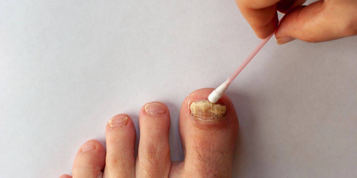
What To Do When Your Toenail Is Falling Off
Read More
Listen Now
What To Do When Your Toenail Is Falling Off
Read More
-
 Listen Now
Flip-flops Causing You Pain? Protect Your Feet This Summer!
Read More
Listen Now
Flip-flops Causing You Pain? Protect Your Feet This Summer!
Read More
-
 Listen Now
Could Feet Be the Windows to Your Health?
Read More
Listen Now
Could Feet Be the Windows to Your Health?
Read More
-
 Listen Now
Why Are My Feet Different Sizes? It's More Common Than You Think
Read More
Listen Now
Why Are My Feet Different Sizes? It's More Common Than You Think
Read More
-
 Listen Now
Revealing the Secrets of Men's and Women's Shoe Sizes: Why Are They Different?
Read More
Listen Now
Revealing the Secrets of Men's and Women's Shoe Sizes: Why Are They Different?
Read More
-
 Listen Now
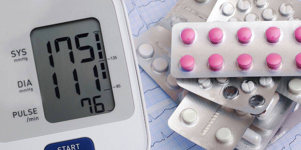
Do Blood Pressure Medicines Cause Foot Pain?
Read More
Listen Now
Do Blood Pressure Medicines Cause Foot Pain?
Read More
-
 Listen Now
15 Summer Foot Care Tips to Put Your Best Feet Forward
Read More
Listen Now
15 Summer Foot Care Tips to Put Your Best Feet Forward
Read More
-
 Listen Now
What Is Erythromelalgia?
Read More
Listen Now
What Is Erythromelalgia?
Read More
-
 Listen Now
What Are Shin Splints?
Read More
Listen Now
What Are Shin Splints?
Read More
-
 Listen Now
Is Foot Analysis Better than Horoscopes? What Do Your Toes Reveal About Your Personality?
Read More
Listen Now
Is Foot Analysis Better than Horoscopes? What Do Your Toes Reveal About Your Personality?
Read More
-
 Listen Now
Swollen Feet During Pregnancy
Read More
Listen Now
Swollen Feet During Pregnancy
Read More
-
 Listen Now
Custom Orthotics vs. Over-the-Counter Inserts: Which Are Best for Your Feet?
Read More
Listen Now
Custom Orthotics vs. Over-the-Counter Inserts: Which Are Best for Your Feet?
Read More
-
 Listen Now
How Many Steps Do I Need A Day?
Read More
Listen Now
How Many Steps Do I Need A Day?
Read More
-
 Listen Now
How To Tell If You Have Wide Feet
Read More
Listen Now
How To Tell If You Have Wide Feet
Read More


