- Home
- Foot & Ankle Conditions
- Morton's Neuroma
Morton's Neuroma: causes, symptoms and treatments
What is Morton's neuroma?
A neuroma is a benign growth that involves thickening and inflammation of nerve tissue that results in pain or a burning sensation. A Morton's neuroma is found in the tissue of the toes.
Symptoms of Morton’s neuroma include pain in the ball of the foot, feeling like you’re standing on a pebble, burning pain, foot pain or numbness.
- What are common causes of Morton's neuroma?
- What are common risk factors for Morton's neuroma?
- How is Morton's neuroma diagnosed?
- Nonsurgical treatments for Morton's neuroma
- Advanced Morton's neuroma treatment options
- Why UFAI is the best choice for Morton's neuroma care?
- Morton's neuroma FAQs
- What kind of doctor treats Morton's neuroma?
- Can Morton's neuroma cause leg or calf pain?
- What are the best shoes to wear for Morton's neuroma?
- What does Morton's neuroma look like?
- Can Morton neuroma cause sciatica?
- Can Morton's neuroma cause numb toes?
- Can Morton's neuroma come back?
- Can an X-ray show Morton's neuroma?
- What can be mistaken for Morton's neuroma?
What are common causes of Morton's neuroma?
A Morton's neuroma usually forms in response to an injury (such as blunt trauma to the forefoot or direct injury to the nerve), overuse, or repetitive motion of the foot and toes. It usually affects only one foot but can occur in both feet.
Over time, the continued compression and irritation of the nerve will cause more severe damage to the nerve. As the damage progresses, conservative treatments become less likely to work. Many patients come to us after other doctors have failed to treat their neuroma pain.
What are common risk factors for Morton's neuroma?
- High heels: Wearing high heels puts pressure on your toes and the ball of your foot.
- High-impact sports: Participating in high-impact athletic activities may subject your feet to repetitive trauma. Sports that feature tight shoes, such as snow skiing or rock climbing, can put pressure on your toes.
- Foot deformities: Patients with bunions, hammertoes, high arches, or flat feet are more prone to developing Morton's neuroma. Altered foot biomechanics associated with these conditions can increase pressure on the forefoot, leading to nerve irritation and compression.
How is Morton's neuroma diagnosed?
Diagnosing Morton’s neuroma requires a medical examination. This involves a discussion of the patient’s symptoms and a review of their medical history and footwear choices. Our foot and ankle specialist will squeeze the two sides of your foot together while putting pressure on the space between your toes. Shooting nerve pain or a “clicking” feeling between the bones can indicate Morton’s neuroma.
In some cases, imaging studies like ultrasound, X-ray, or MRI may be recommended to visualize the neuroma or rule out other causes of foot pain.
A podiatrist's expertise in foot and ankle conditions allows for an accurate diagnosis and the development of an appropriate treatment plan tailored to the individual patient's needs. Often, patients come to us for a second opinion for a neuroma that is not getting better, and we discover they have been misdiagnosed and actually have a plantar plate tear of the toe joint. While they have similar symptoms in the same area of the foot, they are very different conditions with different treatments.
Nonsurgical treatments for Morton's neuroma
Remove the deforming force and stop the activity causing the injury
This is an important first step because if you do not remove the cause(s) of the problem, you are simply treating the pain, while the problem can continue to worsen. If shoes or sports are contributing to your neuroma, you may need to wear shoes with a wider toe-box or to take a break from your sport while it heals.
Immobilization
In some acute cases, immobilizing your foot for a period of time can be very helpful. This is achieved by wearing a special shoe or boot.
Physical therapy
Physical therapy is another very effective treatment for neuromas in their early stages.
Steroid injections
In acute cases, cortisone (corticosteroid injections) can help reduce pain and neuroma symptoms.
Custom orthotics
Custom-molded orthotic shoe inserts are often an important part of treatment and lessen the chance for the neuroma to return.
Over-the-counter medication
Anti-inflammatory medications can reduce swelling and relieve pain.
Advanced Morton's neuroma treatment options
In more severe and chronic cases of Morton’s neuroma that hasn’t responded to conservative therapies, we may need to turn to more advanced treatment. Each of these treatments has unique benefits or downsides, so each of our patients has a customized treatment plan that considers their physical condition and day-to-day personal and professional needs.
Some advanced treatment options for Morton's neuroma are:
Cryotherapy (cryoablation with ultrasound)
This is the method of freezing the nerve. Through a rapid and limited deep-freezing of the affected area, the chronic pain that damaged nerves cause is resolved with a simple and comfortable office procedure. Read more about Cryotherapy.
Alcohol sclerosis
A special alcohol solution is injected over the nerve. This solution has an affinity for nerve tissue and the end result is to deaden the nerve. The loss of the nerve can then leave the patient symptom-free.
This procedure stops the nerve from transmitting nerve signals, and therefore, there is less pain. Most patients respond well to the treatment, which is office-based and done under local anesthesia. In certain cases, the procedure does not fully treat the nerve pain on the first attempt, and a second treatment may be necessary.
Morton's neuroma nerve decompression surgery
One of the most recent advances in neuroma therapy is nerve decompression. In many cases, the overlying ligament that connects the metatarsal heads together is pinching the nerve. A simple surgery releases this ligament allowing for increased space for the nerve and relief of pain. This surgery leaves the nerve intact and allows continued sensation to the toes.
Morton's neuroma nerve excision surgery (neurectomy)
This is the most traditional treatment for neuroma pain. Neuroma surgery involves removing the nerve from the metatarsal head region, which decreases or removes pain. There is a risk of the remaining nerve regrowing or scarring but overall, with proper technique, the risk is minimal.
After years of research and studies done by us and others, our neuroma specialists have learned that burying the nerve into the muscle dramatically reduces stump neuromas and post-surgical problems. A stump neuroma issue is when nerve tissue is severed and tries to regrow. But by burying the nerve in a muscle in the arch area, the nerve is happy, and the chance of regrowth is low.
Why UFAI is the best choice for Morton's neuroma care?
The physicians at UFAI are internationally recognized foot and ankle surgeons. They use the latest technologies and treatment options available in a comfortable and family-friendly environment.
If you or anyone in your family are experiencing foot problems, we’re here to help. Our nationally recognized foot and ankle specialists offer the most advanced podiatric care and the highest success rates nationwide. We are leaders in researching, diagnosing, and treating all foot conditions.
We are conveniently located through the Los Angeles area with locations in or near Santa Monica (on Wilshire Blvd.), Beverly Hills, West Los Angeles, Manhattan Beach, Northridge, Downtown Los Angeles, Westlake Village, Santa Barbara, and Valencia, to name a few.
Morton's neuroma FAQs
What kind of doctor treats Morton's neuroma?
Morton's neuroma is typically treated by a podiatrist. Podiatrists are foot and ankle specialists who perform only foot and ankle care in their practices. The level of training for a podiatrist consists of 4 years of podiatry medical schooling and training, with a specific foot and ankle emphasis. They can provide both non-surgical and surgical treatments if required.
Can Morton's neuroma cause leg or calf pain?
Morton's neuroma can indirectly lead to leg or calf pain. When a person experiences severe pain in the foot, it may alter their gait or walking pattern. This altered gait can cause leg or calf pain.
What are the best shoes to wear for Morton's neuroma?
Look for shoes with
- A wide toe box to give the toes space.
- A low heel to relieve pressure of the ball of the foot.
- Cushioning and support.
- Soft and flexible material.
- Room for orthotics.
- The proper size.
- Laces or adjustable straps for comfort.
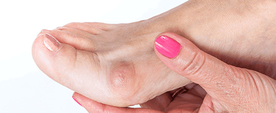
What does Morton's neuroma look like?
A Morton's neuroma typically does not have a visible appearance on the outside of the foot. If you suspect Morton's neuroma and are in Southern California, consult with the doctors at University Foot and Ankle Institute (or a podiatrist near you)
Can Morton neuroma cause sciatica?
Morton's neuroma is not known to cause sciatica. They are distinct conditions that can both lead to foot and leg pain. If you are experiencing symptoms that include foot and/or leg pain, it's important to consult with a healthcare professional.
Can Morton's neuroma cause numb toes?
Yes, Morton's neuroma can cause numbness or tingling in the toes due to compression or irritation of the nerve.
Can Morton's neuroma come back?
Morton's neuroma can recur but not necessarily in the same location. Recurrence can happen for several reasons. If you’ve had Morton's neuroma in the past and are experiencing similar symptoms again, consult with a podiatrist.
Can an X-ray show Morton's neuroma?
X-rays can rule out other conditions, such as fractures or arthritis, but they are not effective for soft tissue problems like Morton's neuroma. For this, your foot and ankle specialist may use ultrasound or magnetic resonance imaging (MRI).
What can be mistaken for Morton's neuroma?
Conditions such as metatarsalgia, capsulitis, stress fracture, bursitis, tarsal tunnel syndrome, neuropathy, and arthritis can be mistaken for Morton's neuroma. It's essential to consult with a podiatrist if you're experiencing foot pain.
-
Foot and Ankle Surgeon and Director of University Foot and Ankle Institute

Dr. Bob Baravaria DPM, FACFAS is a Board-Certified Podiatric Foot and Ankle Specialist. He is an assistant clinical professor at the UCLA School of Medicine and serves as Director of University Foot and Ankle Institute.
Dr. Baravarian has been involved in athletics his entire life and played competitive tennis in high school and college. He has an interest in sports medicine, arthritis therapy, and trauma/reconstructive surgery of the foot and ankle. He is also fluent in five languages (English, French, Spanish, Farsi, and Hebrew),

Learn More from our Morton's Neuroma Blog Articles
 Would highly recommend. Knowledgeable and patient.Robert H.
 Great experience. Great communication. Great direction for my care. Very happy I chose to go with this particular doctor and o...Christopher R.
- I am accident prone and have been using University Foot and Ankle for a number of years. Have seen Dr. Jafary an Dr. Franson. B...Greg K.
- Great service and care. Highly recommend Dr. Franson.David B.
- If you have to go see a Doctor than this is a great experience.Frank M.
- My doctor was great. Really greatRudolph B.
- Good.David E.
- Your Santa Barbara office and Dr. Johnson always give me excellent care!Jayne A.
 Dr Baravarian and the team at UFAI are simply the best. I am 53 and very active. I had surgery to reduce a bone spur that impin...Craig H.
- Dr. Gina Nalbadian was amazing!! I came in with an emergency foot situation and she had wonderful bedside manner and resolved m...Danielle C.
- I was frustrated that after 3 weeks I still hadn’t heard back about my PT referral status. And I did sit in a room for over 30 ...Sarah C.
- I’m very pleased with Dr. Kelman.Alan S.