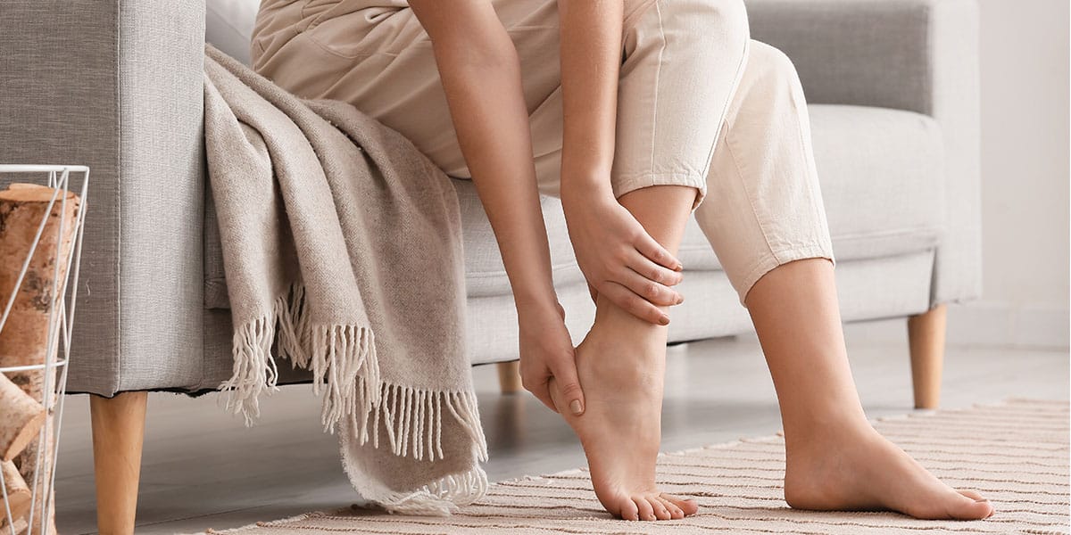
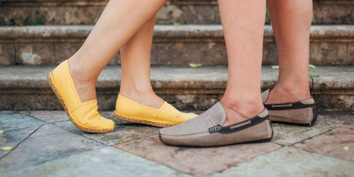
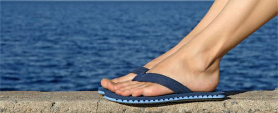
Posted by HubSpot User Default on Thu, Mar 15, 2012 @ 11:01 PM
University Foot and Ankle Insitute and The Los Angeles Ballet: A Perfect Combination


University Foot and Ankle Institute is proud to have been selected by the Los Angeles Ballet Company as the official foot and ankle care center. After years of care for ballet and other dancers, University Foot and Ankle Institute was approached to provide state of the art care for the amazing professional ballet dancers who are part of the Los Angeles Ballet Company.
"We are very proud of this affiliation and feel it allows us to treat these amazing dancers with very high tech. options and offer them the most rapid recovery and the longest sustained careers" noted Dr. David Soomekh, a foot and ankle surgeon at University Foot and Ankle Insitute. Dr. Soomekh has assumed the primary role of caring for the ballet company but is aided by the entire foot and ankle and physical therapy staff at University Foot and Ankle Insitute.
"This is an amazing opportunity for our practice to show how we can care for these elite athletes and also to learn from them how to get patients rehabilitated and back to their profession as rapidly and safely as possible" noted Dr. Bob Baravarian, co-director of University Foot and Ankle Institute.
University Foot and Ankle Institute has been at the forefront of comprehensive foot and ankle care since its inception. The team of doctors and physical therapists care for all types of foot and ankle problems and all age patients with emphasis on compassionate and efficient care. From the most simple to the most complex foot and ankle ailment, no problem is to small or to large to handle.
For an appointment with one of our doctors at one of our 11 Southern California locations please call 877-677-0011 or visit www.footankleinstitute.com
Posted by HubSpot User Default on Thu, Mar 15, 2012 @ 05:39 PM
The Q-Clear Fungus Nail Laser System
University Foot and Ankle Institute is proud to announce the addition of a Q-clear fungus nail laser to our practice locations. The nail lasers will be located at all locations and can be used to treat from the most simple to the most complicated fungus nail cases. The treatment is done in office and is painless. The procedure takes from about 20 minutes to 45 minutes depending on the number of nails involved and the amount of damage to the nails.
Our Fungal Nail Laser Advantage
The Q-clear laser is considered the state of the art laser for fungal nail care and has multiple wavelengths for different nail needs. Unlike first generation nail lasers that did not have multiple wavelengths, the new Q-clear laser allows for adjustments to the amount of depth and heat created and can be customized to the patient’s needs.

University Foot and Ankle Institute:
Advances Fungus Nail Laser Care
With this addition to our practices, we hope that we can continue to provide state of the art care to our patients with integrity and kindness. Although the fungus nail laser is not covered through insurance, we have placed our pricing on a very competitive rate. Unlike other locations, we will be setting pricing by the number of damaged nails and wether one or two feet need treatment. We also have added a fungus nail lacquer and fungus foot cream called PediMD to our treatment regimen that assists in both treating the fungus and helps prevent the fungus from returning after the nail grows out.

For more information or to schedule an appointment please call 877-677-0011.
The fungus nail laser is available in all 11 of our offices including Santa Monica, Beverly Hills, Sherman Oaks, Valencia, Manhattan Beach, Bellflower, Cypress, West Hills,Thousand Oaks and Santa Barbara areas.
Posted by HubSpot User Default on Wed, Feb 08, 2012 @ 11:55 AM
Big Toe Pain: Sesamoid Pathology
The sesamoids are two small round or oval bones situated in the thick soft tissue ligamentous plantar plate under the first metatarsaophalangeal (big toe) joint (Figure 1) and provide attachments for multiple important ligaments and muscle-tendinous units that provide critical stability, strength, and function to the big toe during ambulation and propulsion. They fully ossify by the age of 7. They usually ossify by multiple centers of ossification and rarely this bone can develop an incomplete fusion and bipartide sesamoids have been reported from 7-30% of the time and usually involve the tibial (medial and larger of the two bones) and are usually found to be bilateral. They are infrequently symptomatic however, their tortuous circulation make them prone to slow healing and potential for avascular necrosis when injured.
FIGURE 1 Normal Sesamoids
Type of Sesamoid Injuries
- Sesamoiditis
- Sesamoid fracture
- Sesamoid Stress Fracture
- Symptomatic Bipartide Sesamoids
- Irregular Shaped or Enlarged Sesamoids
- Osteochondritis
- Subluxation or Dislocation of the Sesamoids
Sesamoiditis
Sesamoiditis is defined as inflammation and swelling of the peritendinous structures that inserts around the sesamoid bones. It often occurs in young adults and can be related to acute traumatic event or repetitive weight-bearing load during various exercises. Jumping or falling from a height, unsupportive or high-heeled shoes, excessive activity, or dancing have been reported to cause inflammtion to sesamoid bones. There can be a gradual or sudden onset of pain, swelling, and inflammation to the area und
er the big toe joint worse with or after periods of activity. It is commonly a diagnosis of exclusion because early in the disease process routine foot x-rays will be normal. A bone scan or MRI can be beneficial to aid in the diagnosis. It is important to educate patients that symptoms usually resolve slowly with the below mentioned conservative therapy. If symptoms continue to be painful then surgical excision of the involved sesamoid (Figure 5) may be warranted, however this does cause inherent imbalance of the big toe and deviation can occur.
Symptomatic bipartide fractures, and irregular or enlarged sesamoids usually became painful in a similar fashion and usually respond with conservative fashion. If they fail conservative measures the excision of the involved sesamoid may be warranted. Enlarged or prominent sesamoids can undergo complete excision or “shaving” of the invlolved bone.
Conservative Treatment
- Activity modification
- Supportive shoes
- Metatarsal pads
- Toe strapping
- Stiff soled, rocker bottom shoes with steel shank insert
- RICE
- NSAIDS
- Custom orthotics with pathology specific “cutout” design to decrease pressure under the sesamoids (Figures 3 and 4)

Figure 3 Casting for orthotics Figure 4 Custom Orthotic
Surgical or Invasive options
- Local steroid injections
- PRP injections
- Surgical excision of painful sesamoid (Figure 2)

Figure 5 Surgical excision of sesamoid on x-ray
Sesamoid Fractures
Sesamoid fractures (Figure 6) can develop from acute fracture or secondary to progression or un-treated stress fractures or osteochondritis. Acute fractures including fall from heights, big toe hyper-extension injuries, and other injuries can result in one or multiple fracture fragments of the involved sesamoid. Recurrent micro-trauma such as running, dancing, sporting activities, can cause stress injuries to the bone and compromise circulation resulting in osteochondritis or stress fractures of the sesamoid bones. X-rays can demonstrate acute fractures, however if it is unclear between a bi-partide or sesamoid fracture then MRI and bone scan are beneficial. These pathologies are notorious to slow healing, and usually require an extensive healing period with at least prolonged protective weight bearing in a short leg fracture boot or non-weight bearing in a cast and crutches. It is important to educate patients on the longer recovery with this diagnosis. The transition to a stiff soled shoe and/or custom functional foot orthotic to decrease pressure to the area is required for the following several weeks with activity modification to ensure proper healing. RICE, NSAIDS are encouraged as well. The use of a bone stimulator may be utilized as well in certain cases.
Surgical recommendation is recommended after failure of extensive conservative options as mentioned above. This can include partial excision of the small or large symptomatic fracture fragments or complete excision of the involved sesamoid bone. Again, it should be re-iterated that there are certain inherent muscle imbalances that develops after excision these bones and adjunctive procedures or further surgeries may be in order to maintain a normal position of the big toe.
Figure 6 Sesamoid Fractures shown on these x-rays
University Foot and Ankle Institute
University Foot and Ankle Institute offers comprehensive care for sesamoid pain and fracture. From injection therapy and orthotic care to surgical treatment options, count on our experts to offer you state of the art care in a comfortable and friendly environment.
Posted by HubSpot User Default on Thu, Feb 02, 2012 @ 11:46 AM
Extracorporeal Shock Wave Therapy For Plantar Fasciitis and Achilles Tendonitis
Extracorporeal Shock Wave Therapy or ESWT, was initially used in the treatment of urinary, kidney, and salivary stones. ESWT has been used since the late 1980s for the treatment of various musculoskeletal disorders, including:
- plantar fasciosis/ plantar fasciitis
- Achilles tendinosis/ Achilles tendonitis
- sesamoiditis
- stress fractures
- fresh fractures of bones
- delayed union of fractures
- nonunion of fractures
- wound healing
Currently, we use the treatment for mainly plantar fascitis/fasciosis and for Achilles tendonitis/tendinosis conditions. Although we have used it for would healing, we have found better options for these other conditions.
ESWT is not limited to the foot and ankle problems, as it has also been shown to help:
- patellar tendinopathy (jumper’s knee)
- lateral epicondylitis (tennis elbow)
- medial epicondylitis (golfer’s elbow)
- shoulder calcific tendinitis
Types of ESWT
ESWT has been FDA approved and used in the United States since 2000. ESWT is a non-invas ive treatment that involves the delivery of low- or high-energy shock waves to a specific site to the body. Low-energy shock waves require a series of treatments and do not typically require anesthesia or injections. High-energy shock wave treatment is done in one session and usually requires general or regional anesthesia. Currently, more physicians are using low-energy shock waves without any anesthesia. A recent 86 patient study by Rompe et al. showed that using local anesthesia before shock wave treatments actually reduced the efficacy of ESWT. In another recent study by Klonschinski et al., local anesthetic was found to inhibit and alter the effects of ESWT.
We have used both low energy and high energy shockwave therapy options. With high energy, due to the need to get a good treatment with one shockwave session and the overall cost of multiple sessions, we have found that in certain cases there is an over treatment which may result in stress reactions or bruising of the heel. Furthermore, the treatment is very aggressive and may be overkill. On the contrary, stress fracture or delayed bone healing cases seem to do better with high energy treatment.
Low energy cases seem to be a little less stressful to the foot and do not cause deep bone bruising. Low energy shockwave does require 3 treatments on average, however the treatments can be more catered to the patient’s needs and symptoms.
Mechanism of ESWT
ESWT is thought to work on areas where the body has stopped responding to the area of concern due to the chronicity of the problem. Multiple studies have shown that over time, there is a reduction in the amount of blood flow to a region of damage. This blood flow is essential for healing of damaged tissue. Without blood flow, there is an increase in chronic scar tissue resulting in chronic pain. ESWT increases trauma to the area causing the return of blood supply which helps with healing. The shock waves travel through fluid and soft tissue and their effect occurs at sites where there is a change in impedance (ie -bone/soft tissue interface). This creates an expansion and compression within the area being treated. The expansion and compression of the soft tissue causes microscopic levels of trauma to the area of concern, stimulating inflammation to the area, thereby promoting blood flow and nutrients to reach the problem area and stimulate healing of the area. It is also thought that ESWT may reduce the transmission of pain signals from the sensory nerves, reducing sensitivity and pain to the foot. It is important that no anti-inflammatory medications are taken after ESWT as it would counteract its purpose.
When do we consider shockwave?
ESWT is considered when patients fail approximately 6 months of conservative therapy. At this point, musculoskeletal problems are generally chronic and require acute inflammatory tissue to resolve the issue. Conservative therapy for musculoskeletal problems often involve:
- icing
- rest
- physical therapy
- custom or over-the counter orthotics
- activity modification
- shoe modification
What is the visit like?
Low-energy ESWT is performed in the office over a period of 3 visits, each spaced about one week apart. Thousands of shock waves are directed at the area of concern without local anesthesia and are custom catered to the patient’s needs. Patients will typically feel mild improvement after the visit, significant improvement after the second treatment and often
are able to return to work and activities of daily life after the third treatment. Patients may be put in a walking boot for comfort or kept in a stiff soled shoe. Some patients may experience bruising, reddening, or swelling of the treated area. The patient should refrain from taking any anti-inflammatory medication so that the effects of the shock wave can be realized without inhibition. Overall, shockwave is a very safe treatment option but there are a few contraindications to shockwave, including bleeding disorders, and pregnancy.
Conclusions
ESWT is similar to treatments such as Platelet-Rich-Plasma, and Topaz ablation therapy in that they turn a chronic, or non-inflammatory, process into an acute, or inflammatory, process, leading to increased blood flow and healing to the area. However, ESWT is the least invasive of the three as it can be done in the office and does not require any needles or opening of the skin. ESWT is becoming one of the mainstays of treatment for many chronic musculoskeletal problems as there is an increasing body of evidence supporting the use of ESWT for various musculoskeletal areas of the body.
Posted by HubSpot User Default on Tue, Jan 17, 2012 @ 08:32 PM
Bellflower and Cypress Podiatry Offices of University Foot and Ankle Insitute Welcome Dr. Doron Nazarian to the Group.
This month's spotlight is on California Podiatric foot and ankle surgeon Dr. Doron Nazarian. After completing a comprehensive foot and ankle residency program at Olympia Medical Center in Los Angeles, Dr. Nazarian completed a comprehensive sports medicine and reconstructive surgery fellowship at University Foot and Ankle Institute.
Bellflower, CA and Cypress, CA Podiatry Locations
Dr. Nazarian can be found at the UFAI Bellflower and Cypress locations serving the residents of Bellflower, Cypress, and also Los-Alamitos, Long beach, Seal beach, La Palma and other regions of north Orange County. The practices are located in the heart of orange county with freeway access from the 91, 405, as well as the 605 freeways. University Foot and Ankle Institute offers the latests technology and surgical expertises in the podiatric medicine, surgery and biomechanics arenas.
Dr. Nazarian concentrates his efforts on common as well as reconstructive foot and ankle pathologies including corrective bunion surgery, pediatric and adult flat feet deformity, sport related injuries and biomechanical abnormalities. Dr. Nazarian is available for consultation at the bellflower and cypress offices. For an appointment, please call 877-677-0011.
Posted by HubSpot User Default on Tue, Jan 17, 2012 @ 08:03 PM
What is a Bunion?
Bunionscome in all shapes and sizes. They are largely genetic resulting from a looseness of the great to and midfoot joints resulting in a shift of the bone and bunion formation. Although genetics are the primary cause, shoes and other factors can accelerate and worsen bunion problems.

Conservative management:
- Well-fitting, supportive shoes
- Custom functional orthotics, stabilizing the medial column of the foot and slowing down the progression of the bunion deformity.
- Bunion night splints
- Padding over the prominent bone
- Toe alignment splints
- Injections and pills don’t play much of a role in managing a painful bunion.
Conservative management of bunion problems are exactly that: management. It is not typically curative or designed to fix the problem. At some point, surgery is recommended. Surgery is usually recommended when the deromity becomes painful, affects shoe choice, and when the discomfort limits activities. Surgery is discouraged if there is no pain, and when cosmesis is the main driving force.
Bunion Surgery Work-up:
Because bunions come in all shapes and sizes, the surgical treatment must include a varied approach.
The choice of bunion surgery depends on:
- The size of the bunion deformity
- The laxity of the bone and how loose the foot bones are
- The angle of the cartilage of the great toe
- Deformity of the great toe bones and/or bunion only
- The length of the metatarsal bone leading to the great toe
Surgical bunion treatment:

-
Simple “bumpectomy”
- This is the simple, minimalist approach. This is the wrong surgery for most people, but can be effective when chosen wisely. Easy recovery with this procedure. Usually not great long term correction, how ever.
. Classic Chevron type osteotomy
- This is the most common procedure done to correct bunion deformities. It includes shaving the prominent bump, but also includes a shift and realignment of the head of the metatarsal and the big toe joint. This is fixated with pins or screws; our choice at UFAI is usually a 2-screw fixation, which is very stable and allows for earlier weight bearing and range of motion. We usually allow weight bearing protected in a walking boot right away after this surgery.

-
Reverdin type osteotomy
- This procedure can correct some angular problems in the region of the deformity, having it’s strongest correction on the joint position/alignment.
-
Akin phalangeal osteotomy
- When there is alignment problems within the toe itself and it is angulated strongly toward the 2nd toe, this procedure removes a wedge near the base of the toe that swings the toe into a straightened position.

-
Base wedge osteotomies
- Though these procedures are considered somewhat unstable, they can be good to correct higher angled bunion deformities. When these are done with wedge bone grafting and screw or plate fixation, they can provide solid correction, still maintain good metatarsal length, and can be relatively stable. Recovery includes a period on crutches, usually about 4-6 weeks.

-
Lapidus procedure (wedge corrective fusion of the 1stmetatarsal cuneiform joint)
- This procedure gets to the root of many bunion pr oblems. By correcting and stabilizing this joint at the base, strong correction can be achieved. This usually affords excellent long term correction. This is usually the procedure of choice for younger people with a “hypermobile” midfoot joint. This requires cast and crutches for 4-6 weeks or so after surgery.
The University Foot & Ankle Advantage:
Bunion Surgery has become more refined over the years. At the University Foot & Ankle Institute, we custom-design the bunion surgical procedure and experience for the individual. Every foot and circumstance can be unique an no one procedure is well suited to all foot types. At University Foot and Ankle Institute, we are flexible in using the appropriate combination of procedures to achieve quick recoveries as well as the best long-term outcome.
Posted by HubSpot User Default on Tue, Jan 17, 2012 @ 07:49 PM
Achilles Tendon Problems? Treatment Options are Available!
The Achilles tendon is the largest and strongest tendon in the human body. It is crucial in transmitting forces responsible for walking, running and various other activities. An injury or tear within the Achilles tendon can cause substantial pain and limitation due to its importance in these daily activities.
Anatomy:
The Achilles tendon is made up of the tendons of the gastrocnemius muscle and soleus muscle, which make up the largest muscles within the calf. It inserts at the back of the calcaneus (heel bone) and many of the tendon fibers can extend further underneath the heel. Tears of the Achilles tendon can be partial tears, which only involve a portion of the tendon or complete tears. Most often a tear occurs in the mid-portion of the tendon, often referred to as the “water shed” area due to its limited blood supply. It is the limited blood supply that can make Achilles tendon tears challenging and can take an extended length of time to heal.
Etiology:
Tears of the tendon are often a result of indirect trauma. Overload forces exceed the tensile strength of the tendon which results in tearing of the tendon fibers. Complete ruptures most often occur in individuals between 30 and 45 years of age with the majority of injuries sustained during sporting activities. Tendon tears are can also in patients with systemic diseases such as rheumatoid arthritis, lupus, chronic hemodialysis and many others. Other factors correlated with potential Achilles tendon tears are steroid use, fluoroquinolone antibiotics and previous injury to the tendon.
Symptoms of Torn Achilles Tendon:
- Pain
- Swelling
- Bruising
- Loss of strength
- Palpable gap in tendon
Diagnosis:
The most crucial step to diagnosis of Achilles tendon tears is a thorough and detailed physical examination. Patients may not experience complete loss of strength in many instances. Because of this and the swelling in the area, up to 20% may be missed on initial presentation. A delay in treatment can have long lasting effects on the overall outcome and thus emphasizes the importance of being evaluated by a foot and ankle physician in a timely manner.

Radiographs may be taken to rule out other associated injuries. To fully evaluate the extent of Achilles tendon injury, an ultrasound, or more often, an MRI will be performed. This allows the physician to visualize the extent of tendon injury and determine the best treatment plan.
Treatment:
Treatment will vary depending on the extent of tendon tear, duration the tear has been present, and the patient. An acute, complete rupture of the tendon requires surgical intervention. This is typically done by re-approximating the torn ends of the tendon to allow for appropriate healing. For the best function results, the repair should occur within one week in order to avoid retraction of the tendon ends. Unless the patient is relatively sedentary, cast immobilization as the primary treatment of an Achilles tendon rupture should be avoided due to the increased risk of re-rupture, decreased strength and sub-optimal functional results.

Achilles tendon tears that are not treated for longer than 4 weeks are considered chronic. Because of the delay in treatment, the tendon ends are become retracted and cannot be repair like an acute rupture. Chronic Achilles tendon ruptures are typically repaired surgically and involve more extensive tendon transfers, flaps or grafts. After surgical intervention, patients are immobilized in a cast for 2-3 weeks, followed by a transition to a walking boot. It is essential to try to begin strengthening of the tendon as quickly as possible to avoid weakening. This needs to be done carefully by trained physical therapist who understand the recovery protocol of Achilles ruptures.
The University Foot and Ankle Institute Advantage
At University Foot and Ankle Institute, we understand the complete function and necessary recovery needs of Achilles tendon ruptures. We pride ourselves in performing state of the art surgery for Achilles tears and we also have added several new designs to the surgical repair procedure to improve and speed up recovery including stem cell grafting and soft tissue augmentation procedures for added strength. Finally, through our on site physical therapy services, we are able to provide rapid and tested recovery protocols to return you to full function in a timely and safe manner.
Posted by HubSpot User Default on Tue, Dec 27, 2011 @ 04:06 PM
Advancement in Achilles Tendon Tear and Achilles Rupture Care
The Achilles tendon is the strongest tendon in the body but it is also the one that is most commonly injured. In this article we discuss the anatomy of the Achilles tendon as well as give an overview on Achilles tendon ruptures.
Achilles Tendon Anatomy
The Achilles tendon is approximately 15 cm in length and is made up of a combination of fibers that are derived from two calf muscles which twist 90o and insert at the back of the calcaneus or heel bone. The Achilles tendon gets its blood supply from 3 sources. There is an area on the Achilles tendon approximately 2 to 6 cm above its insertion to the calcaneus that has poor circulation and is called the watershed region. Due to the diminished blood supply, this is a common area for Achilles tendon lesions and ruptures.

Acute Achilles tendon ruptures
Achilles tendon ruptures occur most often in middle-aged men during athletic activities. Patients with Achilles tendon ruptures often say that they felt a sudden pop or snap in their calf with subsequent weakness or difficulty with ambulation. Some say they felt someone kicked them or hit them from behind but there is noone in the vicinity. Patients may be able to walk with minor swelling and pain after a tear but have weakness.
Achilles Tendon Tear Exam
On examination, a palpable defect in the tendon can be felt and there is often an increase in upward movement of the ankle due to the lack of tension from the Achilles tendon. A common test that is used to evaluate the integrity of the Achilles tendon is the Thompson calf-squeeze test. Squeezing the calf muscles on the affected side will yield little to no movement in the ankle when compared to the unaffected side.
Imaging of Achilles Tendon Tears or Ruptures
MRI and ultrasound are often used to confirm the presence of an Achilles rupture and to evaluate the extent of the rupture. The diagnosis of Achilles tendon ruptures, however, is based on clinical examination. Imaging is often used for surgical planning or in cases in which there are equivocal examination findings.
Achilles Tendon Tear Surgical Advancements
Operative and nonoperative methods offered by foot and ankle doctors have shown favorable outcomes. Generally with surgical repair, there have been reports that there is a decreased rate of re-rupturing the tendon, improved strength, improved ankle motion, better return to activities, and fewer complaints.
Operative treatment involves reapproximating the two ends of the ruptured tendon together using special suture techniques. A repair can be augmented with a tendon transfer, or synthetic graft. Rarely, the Achilles tendon may rupture at its insertion into the calcaneus. In these cases, a tendon anchor may be used to reattach the Achilles to the bone. The patient is then put in a non-weightbearing cast for 4 to 6 weeks followed by gradual weight bearing and physical therapy.
University Foot and Ankle Institute Achilles Tendon Tear Advancements
University Foot and Ankle Institute has performed extensive surgical research into the ideal treatment and rehabilitation of Achilles tendon tears and ruptures. By using a very strong stitching material, often augmenting the tear with a tendon wrap, and getting a very strong repair of the tendon, rehabilitation and cast removed is done rapidly. By not casting the patient for a long time, the Achilles does not lose as much strength and return to activity is quicker. University Foot and Ankle Institute is proud to offer the latest advancements in the repair of Achilles tendon tears and Achilles ruptures.
Posted by HubSpot User Default on Wed, Aug 17, 2011 @ 10:24 AM
The STAR and Inbone Total Ankle Replacements: Which is best for Ankle Arthritis Cases

With the advent of new technology and an improved biomechanic understanding of ankle function, the treatment of ankle arthritis has broadly changed in the past decade. Ankle replacement is rapidly becoming a comparable option to ankle fusion in the treatment of ankle arthritis. The new generation of ankle replacements have provided an excellent option in the treatment of ankle arthritis and have proven to be a better option to ankle fusion in the proper situations. The purpose of this article is to provide the current options for ankle replacement and also to educate the reader to the benefits and current thinking for the use of the different ankle replacements available on the market today. It is important to remember that for the best potential outcome, patient selection and proper placement of the implant is key to an ideal outcome.
Ankle Replacement Patient Selection:
Ankle replacement procedures are outstanding as long as the proper implant is placed in the proper patient. Furthermore, proper patient selection for replacement versus fusion procedures is essential for the best outcome. In general, ankle fusion is reserved at our institute for younger very active patients who require a great deal of strength and stability from their ankle. Ankle fusions are also preferred for cases of severe angular deformities which may not be correctable with ankle replacement. If the ankle is not well aligned and will not become well aligned with boney or soft tissue procedures prior to placement of the ankle replacement implant, an ankle fusion is often performed. Ankle replacement is reserved for patients who do not perform heavy manual labor, are over the age of 45 and preferably over the age of 55 and have little to no ankle deformity. Although some level of ankle deformity may be corrected with soft tissue or bone realignment procedures, the ankle replacement patient of choice is one with an arthritic ankle that is well aligned. Of interest is the fact that patients who have arthritic midfoot and hindfoot joints about the ankle seem to do better with an ankle replacement than an ankle fusion. This is due to the fact that an ankle fusion places greater stress on the surrounding joints which may lead to further arthritic changes of the hinfoot joints, while an ankle replacement allows motion at the ankle resulting in less strain at the surrounding joints.
Ankle Replacement Pre-operative Planning:
Patients will often present to our offices with a pre-conceived notion of what procedure is best for them. This is important to address early in the relationship between the patient and the doctor. It is important to explain the risks and benefits of replacement verus fusion to the patient and to also explain the two procedures in detail. A complete examination of the vascular and neurologic status are undertaken. If the circulation of the patient is poor, an ankle replacement is not selected at the anterior incision and soft tissue complications associated with ankle replacement may not be in the patient’s best interest. Furthermore, a neuropathic patient with lack of sensation to the foot is not a good ankle replacement candidate and such cases have shown an increased rate of failure. The dermatolic work-up require adequate check of the skin for breakdown and quality. If the skin is of poor quality, a skin graft or flap has been placed on the anterior ankle or if there are severe varicocities noted, a possible contraindication to ankle replacement may be noted. Finally, the function of the muscles, ligaments, tendons and bones of the foot and leg must be checked prior to consideration for an ankle replacement. Laxity about the ankle from previous ligament injury must be addressed prior to ankle replacement to build a solid platform for the replacement. A non functioning tendon must also be considered to make sure the pull about the ankle is ideal. Finally, alignment of the bones must be ideal or be corrected prior to ankle replacement. In most cases, standard radiographs are enough to address a potential ankle replacement. Alignment and bone stock is checked as is the level of arthritis at the ankle and surrounding joints. If there is a potential for cystic changes in the talus or tibia or signs of avascular necrosis, an MRI or CT scan may be ordered to further assess the ankle joint. In the case of avascular necrosis, as long as the majority of the talus is okay, an ankle replacement may be undertaken but if the talus is very damaged, an ankle fusion may be a better option. If there is severe arthritic changes of the joints surrounding the ankle, a potential hindfoot fusion and ankle replacement may need to be performed.
The STAR Mobile Bearing Versus The Inbone Fixed Ankle Replacement Options:
Currently, our institute utilizes two different ankle replacement options. The InBone total ankle replacement (Wright Medical) is a fixed ankle replacement while the STAR total ankle replacement (Small Bone Innovations) is a mobile bearing option. Both have shown excellent results and both have pros and cons. The main different between a fixed and a mobile bearing ankle replacement is that in the mobile bearing option, the plastic spacer material is not attached to the talar or tibial component and floats in between the two. This allows for some varus and valgus tilt and it is suggested that there is less stress during movement on the metal-bone attachment point of the implant resulting in less loosening. The fixed option has a plastic spacer that is attached to the tibial component which does not allow varus or valgus tilt but allows for a long stem insertion into the tibia which has been suggested to add strength and prevent subsidence. The choice of implant is a difficult one to make. The STAR implant is very low profile and has a excellent potential for conversion to either a fusion or larger ankle replacement in the case of a problem. On the other hand the InBone option has an excellent implantation guidance system and the long tibial stem allows for excellent tibial stabilization. Both systems have had excellent results and are fairly easy to insert after a learning curve is mastered.
In our institute, we have began to use the STAR ankle replacement in stable and simple ankle replacement cases which require minimal soft tissue or boney procedures. The Star also has a more stable talar component which we prefer in cases of severe talus arthritic changes or severe medial and lateral gutter arthritis. It is still unclear if there is a major benefit to a three piece mobile bearing implant. On the other hand, if there is any cystic changes in the tibia or if there is previous distal tibial fracture and the bone is not ideal, we prefer the InBone ankle replacement as it has a far more stable tibial component. Also, the InBone ankle replacement has a better surgical guidance system which allows better positioning in more difficult ankles. Finally, in the case of a previously failed ankle replacement that requires revision, the InBone is our preferred system.
Ankle Arthritis Treatment Options Conclusions:
In conclusion, patient selection and proper surgical procedure are essential for ideal outcomes with ankle replacements. With the continued advancements in ankle replacement options, further clarifications will be made to which procedure and replacement material is best for each type of patient. In general, we prefer the mobile bearing ankle in straight forward cases or cases with severe talar damage or medial/lateral gutter arthritis. We prefer the more stable fixed bearing option for tibial cystic changes, deformed tibias, revision cases or more difficult alignment cases. Both replacement have worked very well and have proven to be an excellent addition to our practice with many happy patients.
The University Foot and Ankle Institute Advantage:
University Foot and Ankle Institute is proud to have established a comprehensive ankle arthritis treatment center. From conservative injection therapy and bracing to the most complicated ankle replacement and fusion options, there is no procedure to small or large to handle. Our 9 podiatric foot and ankle surgeons work in a team setting to deal with ankle arthritis cases in a timely and advanced manner utilizing the latest proven technology.
Posted by HubSpot User Default on Thu, Aug 11, 2011 @ 06:57 AM
Lisfranc Mid Foot Fracture Dislocation
There are many different types of fractures that can occur in the foot. Most foot fractures are cause by an external force acting on the foot that will fracture or break the bone. There are a group of joints in the middle of the foot that are situated behind the long bones (metatarsals) of the foot. Collectively these joints are called the tarsometatarsal joint or Lifranc’s Joint.
When there is a forced twisting of the foot, like in a fall, and the middle of the foot is asked to move in a direction it can not, this Lisfranc’s joint can become disrupted and or fractured. First, the ligaments that hold the 2nd metatarsal to the midfoot bone that lies behind the 1st metatarsal next to it (medial cunieform). This ligament will often tear and allow a gapping between these bones. Once this happens, the 2nd metatarsal is now misaligned from the bone behind it (middle cunieform). This misalignment will eventually lead to erosion of the joint and arthritis and pain. This type of foot fracture or dislocation is often misdiagnosed as a simple sprain of the foot. A more severe injury as the middle of the foot continues to twist will then push the the rest of the metatarsals and they can then all slide off the bones behind them causing a more severe dislocation. If there is severe misalignment of several major joints, that if not corrected, will lead to arthritis and severe pain.
Lisfranc Foot Fracture Injury Diagnosis
Lisfranc injuries are diagnosed in several ways. When there is a high suspicion for a midfoot sprain, a detailed history and clinical examination can help to confirm the evidence of the injury. The patient usually presents with pain along the 1st and 2nd rays, with pinpoint palpation pain within the space between the bases of the 1st and 2nd metatarsals and the medial cuneiform. There is often pain in this same area with simultaneous medial and lateral compression or squeezing of the midfoot from side to side. The experienced clinician should be able appreciate the instability of the 1st metatarsal complex through the examination and range of motion. Weight bearing x-rays should be used to determine the degree of injury, including stress views. In those cases where there is a suspicion on plain x-rays, an MRI or CT will be indicated and helpful in the final diagnosis of the severity of the sprain or foot injury.
Subtle Lisfranc Foot Fracture Injury
With most of the slight or minimal sprains of this joint, the foot can be stabilized with a non weight-bearing cast for a period of 4 to 6 weeks followed by a walking boot and several weeks of physical therapy. In many cases, however, surgery is indicated. It is imperative to realign the bones with one another in an anatomical fashiion.
This can only be achieved by surgical reduction and correction of the injury. In the case of the more subtle ligament tear and slight dislocation, the 2nd metatarsal must be brought back to position by buttressing it to the medial cunieform, the bone behind the 1st metatarsal. This can be achieved by using a screw or special strong stitches that will bind the bones together to the give the ligaments time to heal without undo stress. The stitches are preferred, as the screw would need to be removed later.
Severe Lisfranc Foot Fracture Sprain
In the more severe injury most or all the joints that dislocated would need to be stabilized. This is achieved by placing screws or pins across the joints from one bone to the other through the joint while placing them in alignment. The patients is placed in a cast for 4 to 6 weeks and then placed in a boot for 2 weeks. The screws and pins are then removed. Several weeks of physical therapy will be needed for the foot injury.
Many of the slight sprains of the foot are really more than a simple sprain and are actually dislocations of the middle of the foot and are serious injuries that need special attention. It is imperative that the surgeon recognize this injury and treat it correctly and accordingly.
The University Foot and Ankle Institute Advantage:
The surgeons at the University Foot and Ankle Institute are specifically trained in these types of foot injuries and employ the newest technologies and techniques for realignment and repair of Lisfranc’s foot fracture dislocations. For an appointment in one of our 11 Southern California locations please call 877-677-0011.