- Home
- UFAI in the News
- UFAI Medical Journals
- Cobb Procedure and Tibal Tedon Dysfunction
Cobb Procedure and Tibal Tedon Dysfunction

Written by Bob Baravarian, DPM
Anatomy and Function
The posterior compartment of the leg is divided into superficial and deep group muscles. The superficial group contains the gastrocnemius, plantaris, and soleus muscles. The deep group contains the popliteus, flexor hallucis longus, flexor digitorum longus, and tibialis posterior muscles. The tibial nerve (L4 and L5) innervates all of these muscles.
In the mid-calf area, the tibialis posterior muscle lies between the flexor digitorum longus and flexor hallucis longus muscles. It originates from most of the interosseous membrane, posterior surface of tibia inferior to soleal line, and posterior medial surface of fibula [1]. Distally, its tendon of the posterior tibial muscle lies directly behind the medial malleolus as it courses deep to the flexor retinaculum and superficial to the deltoid ligament. The tendon then provides multiple insertions to different parts of the foot. Its main insertion is into the navicular tuberosity, with extensions to the sustentaculum tali, three cunieforms, cuboid, and bases of the second, third, and fourth metatarsal bones. Deep to the plantar calcaneonavicular (spring) ligament, the tendon may contain an accessory bone called the os tibiale externum, which is found when the ossification center in the navicular tuberosity fails to fuse with the remainder of the navicular [2].
The tibialis posterior muscle functions at the ankle, subtalar, and midtarsal joints. It plantarflexes the ankle while it supinates the subtalar and oblique axis of the midtarsal joints. The posterior tibial tendon is the primary antagonist of the peroneus brevis muscle, which functions as an evertor of the foot. With posterior tibial tendon dysfunction (PTTD), there is a loss of an antagonist force to the peroneus brevis and peroneus longus tendons. Secondary to the chronic and unopposed pull from the peroneal tendons, the heel is forced into a valgus position and there is abduction of the forefoot at the midtarsal joints. With prolonged force and deformity progression, the heel is positioned in a laterally deviated angle, subsequently placing the achilles tendon in a more postero-lateral position at its insertion. The resulting force may ultimately cause a fixed, non-reducible deformity.
Etiology
The pathomechanics of PTTD can be systemic or caused by a single trauma. Holmes and Mann [3] have reported that PTTD has a higher incidence in people with past medical history of hypertension, diabetes mellitus, steroid use, and obesity. Myerson et al. [4] showed significant association of PTTD with psoriasis-associated human leukocyte antigen (HLA) markers. In Myerson’s study, the younger population with PTTD had symptoms of generalized entheso- pathy and systemic manifestations of oral ulcers, colitis, and particularly pso- riasis. Mann [5] also described a high incidence (60%) of os tibiale externum in patients with PTTD. He reported that posterior tibial tendon loses its mechanical advantage when it is inserted mostly at the accessory ossicle.
Frey et al.[6] reported that an area of hypovascularization located posterior and distal to the medial malleolus might be the primary cause for the progression of the deformity. The zone of hypovascularization starts 40 mm from the medial tubercle of the navicular and runs proximally for an average of 14 mm.
Mueller [7] reported four etiologies that lead to PTTD: direct injury to the tendon, pathologic rupture (secondary to systemic disease), idiopathic rupture, and functional rupture, in which the tendon is intact but not func- tioning well.
Recent studies by Brage, Pokharna, and Saroff suggested that a humoral autoimmunity against tendon breakdown products could be the contributing factor for progressive PTTD [8].
Iatrogenic causes due to repetitive use of steroid injections have been reported by Jahss [9]. Inman [10] suggested that rheumatoid arthritis could be a leading factor for a ruptured tendon. Traumatic dislocation of the posterior tibial tendon from minor ankle sprains has also been reported [11]. In conclusion, spontaneous ruptures are more common in tendons with chronic degenerative changes due to a systemic or localized pathology.
Clinical Presentation and Examination
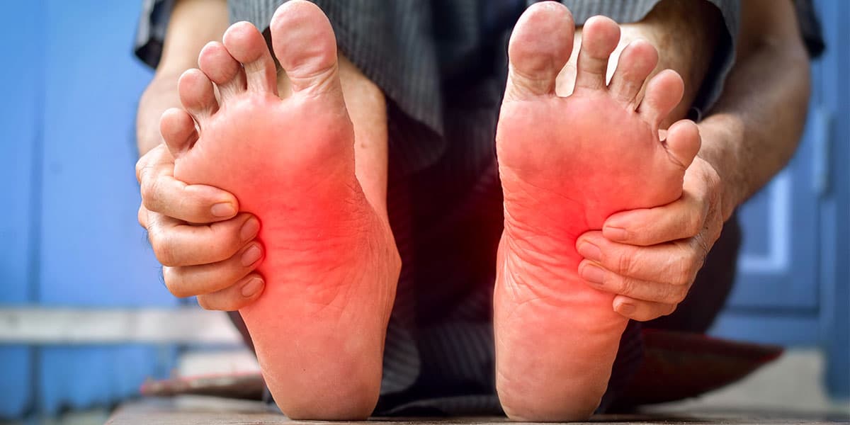
The average patient who presents with PTTD is a female over age forty [12]. Patients often complain of medial ankle or arch pain. The patient often states that her ‘‘arch has fallen.’’ Points of maximum tenderness are found along the course of the tendon, starting at the medial malleolus and progressing to the navicular insertion site. Localized pain, erythema, and edema to the medial aspect of the navicular are also common complaints.
As the deformity progresses, patients will also refer to pain at the lateral side of their foot secondary to excessive pronation. The sinus tarsi area is very tender, due to the valgus rotation of the rearfoot. Over the counter arch supports, strapping, and Nonsteroidal Anti-inflammatory Drugs (NSAID) are remedies that patients might have tried before visiting the doctor. The ‘‘collapsed arch’’ is the chief complaint, and usually is more prominent on the affected extremity than the contralateral unaffected extremity.
Physical examination should be in a very detailed, since the diagnosis can be made clinically and confirmed with imaging modalities. Upon standing, the patient should be given single and double heel-rise tests, and examined for the ‘‘too many toes sign,’’ first metatarsal rise sign, relationship of rearfoot to forefoot, and equinus deformity.
Johnson and Strom [12] have described the ‘‘too many toes sign,’’ in which more than the normal number of one and one-half toes are noted laterally when the affected foot is viewed from the posterior aspect. They have also described the single heel-rise test, in which the patient has difficulty attempting to stand on the affected forefoot secondary to a pathologic posterior tibial tendon. Mann and Thompson [13] have described the double heel-rise test with similar results to bilateral lower extremities. The affected patients were unable to produce calcaneal inversion in cases of PTTD. Hintermann and Gachter [14] proposed a new test called the first metatarsal rise sign. The heel was passively brought into a varus position by externally rotating the shank or grasping the heel itself and rotating internally. The head of the first metatarsal lifted off the ground with PTTD, but remained on the ground with a normal tendon. Their study found that the tests described by Johnson and Strom [12] and Mann and Thompson [13] were negative in 20 to 35% of their study group (21 patients), yet the first metatarsal rise sign was positive in all the cases with PTTD.
The foot should also be observed to determine whether the position of the Achilles tendon on the calcaneus will coincide with the valgus angulation of the calcaneus. The patient is also asked to flex at the ankle with the knee straight and without lifting the heel to check for equinus at the ankle level. The forefoot is viewed from the front for the severity of abduction, medial displacement of the talus, medial arch height, and calcaneofibular impingement at the sinus tarsi area.
In a seated position, manual muscle testing by exerting an abductory force to a supinated foot will determine weakness or possible rupture of the affected tendon [12]. Localized pain upon palpation along the course of the posterior tibialis tendon as well as inflammation and possible erythema at the maximum point of tenderness should help in the diagnosis of PTTD. Equinus is also checked with the knee locked straight and bent ninety degrees.
Differential diagnosis for PTTD should include severe arthritis about the ankle or rearfoot, rearfoot coalition, tibial spurring, tarsal tunnel syndrome, Baxter’s neuritis, plantar fasciitis, tenosynovitis of the tibialis posterior tendon, spring ligament injury, hindfoot fracture, and accessory navicular. The physician should examine for all of the above conditions and order the appropriate tests to confirm the suspected diagnosis of PTTD.
Diagnostic Studies
PTTD is primarily diagnosed by clinical examination. The clinician should perform all the tests described above. On a routine basis, weightbearing radio- graphs are taken to assess the progression of the flatfoot deformity. Radiographic angles including forefoot adductus; cuboid abduction, talar declination, calcaneal inclination, talocalcaneal, and first metatarsal declination should be measured to evaluate the severity of the deformity. Isolated arthritic changes at the midtarsal or hindfoot joints, talonavicular congruency, superimposition of lesser tarsus; medial column breach, tarsal coalitions, accessory bones, and possible fractures should also be assessed by the clinician.
Magnetic resonance imaging (MRI) has the advantage of evaluation for soft- tissue pathology and may also assist in surgical planning (Fig. 2). The tendon is inspected for the extent of the rupture as well as the presence of sheath fluid. Lim et al [15] concluded that secondary MRI signs for posterior tibialis tendon tears included unroofing of the talus, tibial spurring, and tendon sheath fluid. MRI is a confirmatory test that helps determine the extent of the deformity and also aids in the diagnosis of any other related soft-tissue pathology.
Ultrasonography has also been used for the diagnosis of posterior tibial tendon pathology. Miller et al [16] compared 17 preoperative ultrasonograms with their surgical findings and proved that ultrasonography was 100% accurate with the preoperative diagnosis. Further studies by Hsu et al [17] and Chen et al [18] have shown the use of ultrasonography as a valuable and accurate test for the early diagnosis of stage I PTTD and tenosynovitis.
Classification
In 1986, Funk et al [19] classified PTTD based on the location of the tendon rupture: Group I—avulsion at the insertion of the navicular; Group II—rupture within the substance of the tendon; Group III—in-continuity tear of the tendon; and Group IV—tenosynovitis without visible tear of the tendon.
In 1988, Rosenberg et al. [20] classified chronic ruptures of posterior tibialis tendon according to computed tomography (CT) and surgical findings. Type I in- volved an intact but hypertrophied, heterogeneous, low-attenuation tendon, which surgically showed partial ruptures within the tendon substance. Type II involve- ment showed an intact but attenuated tendon. Surgically, involvement was more severe with partial rupture and attenuation of the tendon. Type III tears showed absence of a portion of the tendon, surgically consistent with a complete rupture.
In 1989, Johnson and Strom [12] classified PTTD based on pain symptoms, physical findings, and radiographic changes. PTTD was graded from least to most severe tendon involvement. Abnormalities in foot alignment were also considered in the grading system. Stage I showed peritendinitis or tendon degeneration, mobile and normal alignment of hindfoot, mild to moderate focal pain, mild weakness with single-heel-rise test, synovial tendon degeneration, and normal ‘‘too many toes sign.’’ Stage II involved generalized attenuation of the tendon. Although a mobile and valgus alignment of hindfoot was still present, symptoms included moderate pain along posterior tibialis tendon, marked weakness with single heel-rise test, marked tendon degeneration, and positive ‘‘too many toes sign.’’ Stage III deformity involved a fixed deformity and valgus position of the hindfoot, pain medial and possibly lateral, marked weakness with single heel-rise test, marked tendon degeneration, and positive ‘‘too many toes sign.’’ In 1996, Myerson [8] added a Stage IV, involving the ankle in the overall deformity, to the above classification system. Symptoms include pain at the lateral side of the foot secondary to lateral calcaneofibular impingement syn- drome and eventual lateral tibiotalar degeneration.
In 1991, Mueller [7] also classified PTTD based on increasing severity. Stage I involves an asymptomatic phase with a present biomechanical fault. Stage II shows the beginnings of a symptomatic phase, with symptoms of tendonitis and progressive flatfoot deformity. With progression of symptoms, Stage III is considered a rupture phase, with increasing severity of symptoms, and Stage IV is end stage with rapid progression of the above symptoms severely limiting ambulation.
In 1992, Conti et al. [21] classified PTTD based on MRI findings. Type IA showed one or two longitudinal splits without evidence of intrasubstance degen- eration; Type IB progressed to multiple longitudinal splits and fibrosis without tendon degeneration. Type II dysfunction showed narrowing of the tendon with long longitudinal splits and tendon degeneration. Type IIIA involvement included diffuse tendon swelling with uniform degeneration, and Type IIIB was noted as a complete rupture, with complete replacement of the tendon by scar tissue.
In 1995, Frankel et al. [22] classified PTTD based on clinical, radiographic, and MRI findings, combining Johnson and Strom’s and Mueller’s classifications. Stage I patients showed pain and edema along the tendon, possible weak heel-rise test, relief with rest, and no radiographic signs. Stage II patients had increased pain, minimum relief with rest, medial arch flattening, ‘‘too many toes sign,’’ radiographic signs positive for forefoot abduction, moderate subluxation of the talonavicular joint, decreased arch height, and MRI findings positive for tendon irregularity. Stage III patients had increased forefoot abduction and rearfoot valgus, minimal heel rise, pain at the lateral sinus tarsi, difficulty walking, and MRI findings positive for partial to complete rupture or avulsion of the posterior tibial tendon. Stage IV patients showed end stage findings. These included evidence of rapid progression of degenerative joint disease in the hindfoot on radiographs and severe pain with progression of pes planus. Frankel noted that the only treatment for stage IV diseases was an arthrodesis of the involved joints.
In 2000, Weinraub and Heilala [23] described a new classification scheme for PTTD combining soft-tissue (Stages 1 to 3) and osseous (Grades A to C) pathology, together with a comprehensive surgical treatment plan. This classification is the most detailed and organized of all the systems. It provides differing amounts of damage to the tendon, levels of deformity, and treatment options for each stage/grade combination of deformity. Stage 1 includes acute inflammation, tendonitis, and minimal tendinosis. Stage 2 includes tendinosis with medial attenuation and insufficiency of soft tissue static and dynamic stabilizers, and Stage 3 includes advanced attenuation or rupture of the tendon or medial supportive structures. Osseous deformities are graded as no planar deformity (Grade A), reducible planar deformity (Grade B), or rigid, nonreduc- ible deformity (Grade C). The surgical treatment plan is based on the stage and grade of the deformity. This classification and its associated surgical treatment guidelines are presented in greater detail in another chapter of this periodical.
Conservative Treatment
The type of care needed depends on the patient’s age, past medical history, weight, daily activity level, severity of deformity, and compliance level. Patients that are poor surgical candidates because of their past medical history and daily activity level are best treated with conservative measures. Hypertension, diabetes mellitus, morbid obesity, peripheral vascular disease, and renal and heart disease are some of the problems that may coincide with PTTD [24].
Conservative treatment should also be considered as an initial therapy. A three-month period of RICE (rest, ice, compression, and elevation) therapy may be attempted. Also, immobilization with an Unna boot or below-the-knee casting, and initiation of anti-inflammatory medications should be considered. It may be more appropriate to fit the patient for a removable custom-made foot orthoses or ankle-foot orthotic that depends on the degree of the deformity, the foot type, and the dysfunction stage [25].
Orthopaedic functional shoes, custom-molded shoes, UCBL orthotic, and extrinsic, and intrinsic shoe modifications, including flanged and offset heels with navicular supports, are some recommendations for conservative pedorthic management [25]. Late stages of PTTD with severe deformity may require more dramatic treatment modalities. These include treatment with thermoplastic solid Ankle Foot Orthotic (AFO) and patellar tendon bearing AFO. Appropriate foot- wear is very important for the conservative treatment of PTTD. The purpose of the custom-molded functional orthotic is to reduce hindfoot eversion by placing the subtalar joint in a neutral position and diminishing the length of the posterior tibial complex. The appropriate orthotic device is based on clinical and radio- graphic findings. Initial radiographs should be taken to evaluate any structural deformities and to compare the rearfoot to the forefoot and vice versa.
Steroid injections should be avoided along the medial tendon complex, due to the possibility of weakening or rupture of the medial tendons. If steroid therapy is thought necessary, shot-term oral therapy with protection of the torn tendon may be safer and more helpful to the patient. Lateral sinus tarsi injection may be beneficial in cases of severe arch collapse, yet the treatment needs to be augmented by adjunct orthotic modalities supporting the foot in a more rectus position. Physical therapy may be beneficial during the conservative period of care. Strengthening the posterior tibial muscle, stretching exercises, and ultrasound therapy should be attempted in-patients with early-stage PTTD.
Surgical Treatment
Many surgical procedures have been proposed in the literature for the treatment of adult acquired flatfoot. These include isolated soft tissue and osseous procedures, combination of both osseous and soft tissue procedures, and arthrodesis of the hindfoot or midfoot. Soft-tissue procedures have worked better in early Stage I PTTD, described by Johnson and Strom [12]. Isolated soft- tissue procedures include direct tendon repair with tenosynovectomy, flexor digitorum tendon transfer and spring ligament repair, resection of accessory navicular and transposition of posterior tibialis tendon under the navicular [26,27], split tibialis anterior tendon transfer, and possible tenosuspension [28] by rerouting the tibialis anterior tendon through a keyhole in the navicular and advancement of posterior tibialis tendon under the navicular. Soft-tissue proce- dures alone might eliminate pain only temporarily, because they fail to address the osseous pathology in the adult-acquired flatfoot deformity. Without added stability in the foot, soft-tissue procedures may fail, due to the underlying deformity. In mild cases of deformity, however, soft-tissue procedures with the addition of orthotic devices may be beneficial.
Inflammatory synovium can cause tendon degeneration and lead to a partial or complete rupture if not treated appropriately [29]. Early treatment with tenosy- novectomy plays a critical role in the prevention of tendon degeneration. Crates and Richardson [30] treated seven patients with a Stage I PTTD by performing tenosynovectomy with or without tendon debridement. Six out of seven patients were pain free in an eleven-month follow-up. Johnson and Strom [12] also had satisfactory results with tenosynovectomy after conservative care failed in patients with Stage I PTTD.
Flexor digitorum tendon transfer is the most commonly used tendon transfer for the treatment of posterior tibial tendon dysfunction. However, a second tendon transfer may also be used. With the Cobb procedure, the medial half of the anterior tibial tendon is transferred through a drilled hole in the medial cuneiform or navicular, and a reconstruction of the posterior tibial tendon is performed. In 1990, Helal [31] described satisfactory results in five out of eight patients with use of the split tibialis anterior tendon procedure. In 1998, Weil et al [32] reported excellent results using the Cobb procedure in patients having a Stage 2 PTTD deformity according to Mueller’s developmental stages. The goal of the Cobb procedure is to reconstruct a functional posterior tibial muscle associated with a torn posterior tibial tendon. The Cobb procedure is not considered a functional transfer, and relies on the function of the posterior tibial tendon for its action. Although the procedure has been performed as an exclusive procedure with good results, in most cases the procedure should be combined with other soft-tissue and osseous procedures that will reposition the foot, decreasing the stress on the reconstructed tendon complex.
The osseous procedures commonly used can be divided into two categories: Category A comprises calcaneal osteotomies, medial column stabilization proce- dures, and lateral column lengthening procedure; and Category B includes arthrodesis at the talonavicular joint, Chopart’s joint, subtalar joint, or triple arthrodesis. Category A osseous procedures are often accompanied by tendon transfer and tendoachilles lengthening or gastrocnemius recession. Soft-tissue procedures are usually not indicated in Category B osseous procedures [33]; however, recent studies by Johnson et al [34] have shown excellent correction of hindfoot valgus and forefoot abduction through STJ fusion combined with Flexo- digitorum longus (FDL) transfer and spring ligament repair. The equinus deformity often associated with PTTD should always be addressed with either a soft-tissue or an osseous procedure.
Category A osseous procedures should be selected according to clinical and radiographic findings of the primary plane of compensation associated with planal dominance. Frontal plane deformities are usually addressed by calcaneal osteoto- mies. Medial displacement calcaneal osteotomies direct the gastrocnemius-soleus complex into a more medial position and decrease the valgus force on the calcaneus. In 1893, Gleich [35] was the first to perform a medial displacement of the calcaneus to correct for the hindfoot valgus. In 1923, Lord [36] used a similar calcaneal osteotomy by displacing the calcaneus anteriorly, inferiorly and medially. In 1946, Chambers [37] described the concept of extra-articular calcaneal osteotomy by placing a bone graft under the sinus tarsi to decrease lateral displacement of the talus on the calcaneus. In 1959, Dwyer [38] suggested a lateral opening wedge calcaneal osteotomy with the use of bone graft, and in 1960 [39], he described a closing wedge osteotomy from the medial side of the heel. In 1964, Baker and Hill [40] described the insertion of bone graft into an osteotomy performed inferior to the posterior facet of the subtalar joint. In 1967 [41] and 1974 [42], Silver et al. reported excellent results with lateral opening wedge osteotomy and the use of allogenic bone graft in cerebral palsy patients. In 1971, Koutsogiannis [43] described an oblique medial displacement of the calcaneus for the correction of heel valgus. In 1973, Selakovich [44] described an opening wedge osteotomy of the sustentaculum tali with an insertion of bone graft, along with tightening of the spring ligament, advancement of the posterior tibialis tendon, and transfer of tibialis anterior tendon into the navicular. In 1991, Jacobs and Geistler [45] described a crescentic calcaneal osteotomy for the medial and plantar displacement of the heel. In 1995, Myerson et al [46] combined an FDL transfer with a medial displacement calcaneal osteotomy, and showed significant improvement in radiographic measurements. Catanzariti et al. [47] showed similar results by performing a posterior calcaneal displacement osteotomy (PCDO) and different ancillary procedures, with the FDL transfer being the most common.
Transverse plane dominant pes plano-valgus deformity is best addressed by lateral column lengthening procedures. In 1975, Evans [48] corrected the forefoot abduction with an opening wedge osteotomy and insertion of bone graft in the distal anterior aspect of the calcaneus. The goal of this procedure is to lengthen the lateral column and realign the midtarsal joint. In 1997, Cooper et al [49] reported excessive pressures in the calcaneocuboid joint with an Evans proce- dure. More recent lateral column lengthening procedures include calcaneocuboid distraction arthrodesis for the treatment of the adult acquired flatfoot [50]. The goal of the calcaneocuboid fusion is to avoid the increased pressure in the joint associated with the Evans procedure.
Sagittal plane dominant pes plano valgus deformity is best addressed by medial column stabilization procedures. In 1923, Lowman [51] was the first to describe medial column fusions by a talonavicular fusion, tendoAchilles length- ening (TAL), rerouting the tibialis anterior under the navicular into the spring ligament and using the medial aspect of the Achilles tendon as an accessory ligament tenodesed to the medial arch. In 1927, Miller [52] described fusion of the navicular-cuneiform-first metatarsal joints. A TAL and posterior tibial tendon and spring ligament advancement were also performed. In 1931, Hoke [24] described the fusion of the navicular to the first and second cuneiforms along with the TAL.
A fundamental element in dealing with PTTD is understanding the primary plane of compensation of the adult-acquired flatfoot deformity in order to select the appropriate procedures. Category A osseous procedures are now accompanied by soft-tissue procedures. Double calcaneal osteotomy (PCDO and Evans pro- cedure) with FDL transfer showed great results by Frankel et al [22]. In 1997, Pomeroy and Manoli [53] reviewed 20 cases of Stage 2 PTTD that were treated by TAL, FDL to medial cuneiform transfer, lateral column lengthening, and medial displacement calcaneal osteotomy. Their results showed significant radiographic and clinical improvement. In 1998,Weil et al [32] combined a lateral column lengthening (Evans) and Cobb procedure for the treatment of Stage 3 deformity according to Mueller’s developmental stages of PTTD. He reported great radio- graphic improvement, but 50% of the patients complained of lateral column pain postoperatively. In 1999, Sangeorzan et al [54] combined medial column stabili- zation and lateral column lengthening procedures with significant radiographic improvement. All of his procedures included FDL transfer to the medial cuneiform or stump of posterior tibialis tendon and TAL or gastrocnemius recession.
Category B osseous procedures are usually reserved for the severe arthritic changes or fixed deformity in the midfoot and hindfoot. Isolated, double, and triple arthrodesis sites have been described in the literature. In 1999, Harper [33] reported achieving an 86% patient satisfaction rate by performing an isolated talonavicular arthrodesis. In his series, three patients had an additional FDL transfer to the medial cuneiform, but he found it unnecessary for the support in the medial longitudinal arch. In 1997, Kitaoka and Patzer [55] studied 21 patients who were treated with STJ fusion for PTTD. Satisfactory results were reported in 16 out of 21 patients. Stephens et al [56] in 1999 suggested a repositional STJ arthrodesis to produce excellent correction in a severe flatfoot deformity. In 1994, Clain and Baxter [57] reported the use of simultaneous calcaneocuboid (C-C) and talonavicular (T-N) fusion in different clinical cases with 75% satisfactory results. In 1999, Mann and Beaman [58] reported the results of 24 double (calcaneocu- boid and talonavicular) arthrodeses with 83% overall satisfactory results. Indi- cations for the double arthrodeses included forefoot varus greater than fifteen degrees and mobile subtalar joint free from arthritic changes.
Both isolated and double arthrodesis procedures have been shown to produce compensatory arthrosis in adjacent joints. An alternative to most surgeons is the triple (C-C, T-N and STJ) arthrodesis procedure. In 1993, Sangeorzan et al. [54] reported satisfactory results with triple arthrodesis in 34 out of 40 patients. Ra- diographic measurements including lateral talometatarsal, lateral talocalcaneal, and Anterior-posterior (AP) talometatarsal angles were significantly improved. In 1999, Fortin and Walling [59] also reported good results with triple arthrodesis in 31 out of 32 patients.
The Cobb Procedure
The authors have found that the Cobb procedure is not ideal for all patients. If the posterior tibial muscle is found to be non-functional or severely scarred due to prolonged disuse, an active tendon transfer is preferred. Therefore, the patient is consented for either a flexor digitorum longus tendon transfer or a split anterior tibial tendon transfer. The posterior tibial tendon is identified through an 8 cm curvilinear incision extending from the posterior aspect of the medial malleolus to the medial aspect of the navicular-medial cuneiform articulation. All scarring is freed from the proximal tendon by using the index finger in a sweeping circumferential rotation about the proximal aspect of the incision. The muscle belly is then pulled distally and if found to be low lying, stimulated with the use of a bovie. If there is strong twitch of the posterior tibial muscle, a Cobb procedure is attempted. In most cases, the muscle is not able to be drawn into the distal incision.
In such cases, a decision is made on using a split anterior tibial tendon transfer, based on the amount of scarring associated with the tendon. If the tendon can be freely pulled distally after all scarring is freed, then the procedure may be used. The tendon is checked in a circumferential fashion and all abnormal portions are debrided. Only in cases of severe attenuation or destruction should an entire section of the tendon removed. Following a complete debridement of the posterior tibial tendon, a 2 cm incision is made at the musculo-tendinous junction of the anterior tibial tendon and the medial half of the tendon is identified. The anterior tibial tendon is also identified in the distal incision at its insertion site. After the medial half of the anterior tibial tendon is released proximally, the tendon is harvested through the distal incision, leaving it attached at its insertion.
It is crucial to assure that the tendon is grasped within the tendon sheath through the distal incision before pulling the tendon distally. If this is not done, the tendon may not glide properly at the anterior ankle region and may be difficult to pull distally. Any adjunctive osseous procedures are performed before reconstruction of the posterior tibial tendon, but after harvest of the anterior tibial tendon. The anterior tibial tendon is then routed through a drill hole fashioned in the medial navicular. The drill hole may be facilitated by using the guide wire and drill from a large fragment screw set. In this way, the drill hole may be checked with fluoroscopy after placement of the guide wire, before drilling the navicular hole. While the foot is held in a supinated position, the anterior tibial tendon is woven through the posterior tibial tendon. Distal pull on the posterior tibial tendon and proximal pull on the anterior tibial tendon can tighten the woven tendon complex at the same time. Once adequate tightness is achieved, the two tendons are sewn together. In cases of longitu- dinal tears with central deterioration, the posterior tibial tendon is then closed onto itself after the anterior tibial tendon is placed in the center of the complex. The reconstructed posterior tibial tendon is replaced behind the medial malleolus and its sheath complex is reapproximated.
If the repaired tendon complex is long enough, the tendon should ‘‘pop’’ behind the medial malleolus at the time of relocation. If the tendon easily relocates behind the medial malleolus, it is too loose and the complex must be tightened. Once the tendon complex is relocated posterior to the medial malleolus, the retinaculum, subcutaneous, and skin structures are reapproximated with the surgeon’s preferred suture material. The flexor retinaculum should be reap- proximated loosely in order to avoid constriction of the tendon complex. The patient is then placed in a below-the-knee cast with the foot held in a slightly supinated position.
Postoperative care often depends on adjunctive procedures. If a soft-tissue procedure is done alone, non-weightbearing in a below-knee cast with the foot in slight supination is standard for three weeks. The foot is then placed in a rectus position and a weightbearing below-the-knee cast is used for a second three-week course. At six weeks, the patient is placed in a supportive ankle brace and custom orthotics, and is started in physical therapy for muscle strengthening. Osseous procedures of the calcaneus require six weeks of non-weightbearing followed by three weeks of weightbearing in a below-the-knee walker. Arthrodesis procedures require ten to twelve weeks of non-weightbearing followed by three weeks of guarded weightbearing. In any osseous case, the patient requires physical therapy for muscle strengthening after full osseous union.
Procedure Selections and Preferred Treatments
We use the Cobb procedure for the treatment of Stage 2 and 3 posterior tibial tendon dysfunction. Stage 2 dysfunction is mild to moderate deformity of the rearfoot with no signs of arthritis. There is also normal mobility of the rearfoot joints. In Stage 3 dysfunction, the joints may be arthritic and there is usually a moderate fixed deformity without normal mobility in the rearfoot joints.
Stage 2 cases are well treated with isolated Cobb procedures only if the foot has minimal abnormality and the posterior tibial muscle is strong with minimal tendon degeneration. However, in a majority of cases, Stage 2 deformity is best treated with a combination of the Cobb procedure and osseous reconstruction. We have found both the Evans and medial calaceneal slide osteotomies to have dependably excellent results. In case of an Evans procedure, the osteotomy is spread a distance of 0.75 cm to 1.0 cm. This is enough anterior rotation to reconstruct the abduction of the midtarsal joint. With medial calcaneal slide procedures, the calcaneus is slid 1.0 cm to 1.5 cm medially to reposition the pull of the Achilles tendon. The lateral overhand is impacted to avoid irritation of the sural nerve or peroneal tendons and avoid a calcaneus that is too wide for regular shoe gear. The Cobb procedure is not performed until realignment procedures have been performed. Following completion of osseous procedures, the harvested tendon is routed through the medial navicular and the posterior tibial tendon is reconstructed. A functional posterior tibial tendon without extensive scarring is preferred in osseous cases for proper long-term function. Although the Cobb procedure may act as a static stabilizer, with a non- functional posterior tibial muscle belly the peroneal tendons may begin to overpower this static stabilization, causing a valgus heel position and abduction at the midtarsal joint.
Stage 3 cases of posterior tibial tendon dysfunction may be treated with osseous procedures. However, we prefer to use arthrodesis of the subtalar joint in order to reposition a fixed deformity (Fig. 8A and 8B). Although other arthrodesis procedures may be performed, we have found excellent results, limited delayed or nonunion rate, ease of fixation, and ease of proper positioning with subtalar fusion. Only in cases of severe rearfoot deformity or arthritic changes in the midtarsal joints is a triple arthrodesis performed. The subtalar joint is excellent for repositioning of the heel to the leg; however, the midtarsal joint is still prone to abduction over time from the chronic pull of the peroneal tendons. Therefore, the Cobb procedure is performed to counteract the lateral pull of the peroneal tendons. Although a functional posterior tibial tendon without extensive scarring is preferred, a non-functional static transfer with subtalar fusion is e adequate to avoid the long term deforming forces of the peroneal tendons.
(A) Preoperative lateral radiograph of a patient with posterior tibial tendon dysfunction. Note the collapse in the medial longitudinal arch mainly in the region of the talonavicular joint. (B) Postoperative lateral radiograph following a subtalar fusion. Note restoration of medial longitudinal arch and proper positioning of the talus.
In cases of both Stage 2 and 3 deformity, the underlying equinus deformity is addressed either with an Achilles tendon lengthening or a gastocnemius recession procedure. This is commonly the primary procedure and allows for better positioning of the foot and ankle after completion.
Summary
Numerous surgical procedures have been described for the treatment of the adult acquired flatfoot deformity. The surgeon should review in detail all the clinical, radiographic and imaging tests and propose the best surgical procedure for the patient. Although flexor tendon transfer has shown excellent results, the split anterior tibial tendon transfer is a second option. If used properly, the Cobb procedure results in less functional loss, since only half of the anterior tibial tendon is transferred. Furthermore, the flexor tendon is not disrupted and continues its primary function in the foot and ankle. Ultimately, the goals of the surgical procedure are to alleviate the patient’s symptoms and pain, restore a normal foot alignment, and limit the loss of foot and ankle function without causing any complications.
References
[1] Moore KL. Clinically oriented anatomy. 3rd edition. Williams and Wilkins; 1992. p. 449–458.
[2] Khan MA, Krueger WA, Meetz GD. Lower limb anatomy. UOMHS/CPMS; 1997. p. 19.
[3] Holmes GP, Mann RA. Possible epidemiological factors associated with rupture of posterior tibial tendon. Foot Ankle 1992;13:70–9.
[4] Myerson M, Solomon G, Shereff M. Posterior tibial tendon dysfunction: Its association with seronegative inflammatory disease. Foot Ankle 1986;9:219–25.
[5] Mann RA. Acquired flatfoot in adults. Clin Orthop 1983;181:46–51.
[6] Frey C, Shereff M, Greenidge N. Vascularity of the Posterior Tibial Tendon. J Bone Joint Surg
1990;72A:884 – 8.
[7] Mueller TJ. Acquired flatfoot secondary to tibialis posterior dysfunction: Biomechanical aspects. J Foot Surg 1991;30:2–11.
[8] Mosier SM, Pomeroy G, Manoli II A. Pathoanatomy and etiology of Posterior tendon Dysfunc-
tion. Clin Orthop 1999;365:12–22.
[9] Jahss MH. Spontaneous rupture of the tibialis posterior tendon: clinical findings, tenographic
studies and a new technique of repair. Foot Ankle 1982;3:158–66.
[10] Inman V, editor. Duvries’ surgery of the foot. St. Louis: Mosby CV; 1973. p. 283.
[11] Rolf C, Guntner P, Ekenman I, Turan I. Dislocation of the tibialis posterior tendon: Diagnosis
and treatment. J Foot Ankle Surg 1997;36:63–5.
[12] Johnson KA, Strom DE. Tibialis posterior tendon dysfunction. Clin Orthop 1989;239:196 – 206.
[13] Mann RA, Thompson FM. Rupture of the posterior tibial tendon causing flatfoot. J Bone Joint
Surg 1985;67A:556–61.
[14] Hintermann B, Gachter A. The first metatarsal rise sign: A simple, sensitive sign of tibialis
posterior tendon dysfunction. Foot Ankle 1996;17:236–41.
[15] Lim PS, Schweitzer ME, Deely DM, Wapner KL, Hecht PJ, Treadwell JR, et al. Posterior tibial
tendon dysfunction: Secondary MR signs. Foot Ankle 1997;18:658–63.
[16] Miller SD, Van Holsbeeck M, Boruta PM, Wu KK, Katcherian DA. Ultrasound in the diagnosis
of the posterior tibial tendon pathology. Foot Ankle 1996;17:555–8.
[17] Hsu TC, Wang CL, Wang TG, Chiang IP, Hsieh FJ. Ultrasonographic examination of the
posterior tibial tendon. Foot Ankle 1997;18:34–8.
[18] Chen YJ, Liang SC. Diagnosis efficacy of ultrasonography in stage I posterior tendon dysfunc-
tion: sonographic-surgical correlation. J Ultrasound Med 1997;16:417–23.
[19] Funk DA, Cass JR, Johnson KA. Acquired adult flatfoot secondary to posterior tibial tendon
pathology. J Bone Joint Surg 1986;68A:95–102.
[20] Rosenberg ZS, Cheung Y, Jahss M, Noto A, Norman A, Frey C, et al. Rupture of the Posterior
Tibial Tendon: CT and Surgical findings. Radiology 1988;176:489 – 93.
[21] Conti S, Michelson J, Jahss M. Clinical significance of magnetic resonance imaging in preoper- ative planning for reconstruction of posterior tibial tendon ruptures. Foot Ankle 1992;13:208 – 14.
[22] Frankel JP, Turf RM, Kuzmicki LM. Double calcaneal osteotomy in the treatment of posterior
tibial tendon dysfunction. J Foot Ankle Surg 1995;34:254–61.
[23] Weinraub GM, Heilala MA. Adult flatfoot/posterior tibial tendon dysfunction: Outcomes anal- ysis of surgical treatment utilizing an algorithmic approach. J Foot Ankle Surg 2000;39: 359 – 64.
[24] Hoke M. An operation for the correction of extremely relaxed flatfeet. J Bone Joint Surg 1931; 13:773 – 83.
[25] Steb HS, Marzano R. Conservative management of posterior tibial tendon dysfunction, subtalar joint complex and pes planus deformity. Clin Pod Med and Surg 1999;16:439–51.
[26] Kidner FC. The pre hallux (accessory scaphoid) in its relationship to flatfoot. J Bone Joint Surg 1929;11:831.
[27] Kidner FC. The prehallux in relation to flatfoot. JAMA 1932;101:1539–41.
[28] Young CS. Operative treatment of pes planus. Surg Gynecol Obstet 1939;68:1099–101.
[29] Myerson MS. Adult acquired flatfoot deformity: Treatment of dysfunction of the posterior tibial
tendon. J Bone Joint Surg 1996;78A:780–92.
[30] Crates JM, Richardson EG. Treatment of stage I posterior tibial tendon dysfunction with medial soft tissue procedures. Clin Orthop 1999;365:46–9.
[31] Helal B. Cobb repair for tibialis posterior tendon rupture. J Foot Surg 1990;29:349–52.
[32] Weil Jr. LS, Benton-Weil W, Borrelli AH, Weil Sr. LS. Outcomes for surgical correction for stages 2 and 3 tibialis posterior dysfunction. J Foot Ankle Surg 1998;37:467–71.
[33] Harper MC. Talonavicular arthrodesis for the acquired flatfoot in the adult. Clin Orthop 1999;
365:65 – 8.
[34] Johnson JE, Cohen BE, DiGiovanni BF, Lamdan R. Subtalar arthrodesis with flexor digitorum
longus transfer and spring ligament repair for treatment of posterior tibial tendon insufficiency.
[35] Gleich A. Beitrag zur operativen plattfussbehandlung. Arch Klin Chir 1893;46:358–62.
[36] Lord JP. Correction of extreme flatfoot: value of osteotomy of os calsis and inward displacement of posterior fragment (Gleich operation). JAMA 1923;81:1502–6.
[37] Chambers EF. An operation for the correction of flexible flatfoot of adolescents. West J Surg
Obstet Gyn 1946;54:77–86.
[38] Dwyer FC. Osteotomy of the calcaneum for pes cavus. J Bone Joint Surg 1959;41B:80–6.
[39] Dwyer FC, editor. Osteotomy of the calcaneum in the treatment of grossly everted feet with
special reference to cerebral palsy. In: Hiutieme Congres International de Chirurgie Orthopedique, NY. Bruxelles: Imprimerie Liebens; 1960. p. 892.
[40] Baker LD, Hill LM. Foot alignment in the cerebral palsy patient. J Bone Joint Surg 1964;46A:
1–15.
[41] Silver CM, Simon SD, Spindall E, Litchman HM, Siala M. Calcaneal osteotomy for valgus and
varus deformities of the foot in cerebral palsy: a preliminary report on 27 operations. J Bone
Joint Surg 1967;49A:232–46.
[42] Silver CM, Simon SD, Litchman HM. Long term follow-up observations on calcaneal osteot-
omy. Clin Orthop 1974;99:181–7.
[43] Koutsogiannis E. Treatment of mobile flatfoot by displacement osteotomy of the calcaneus.
J Bone Joint Surg 1971;53B:96–100.
[44] Selakovich W. Medial arch support by operation: sustentaculum tali procedure. Orthop Clin
North Am 1973;4:117–44.
[45] Jacobs AM, Geistler P. Posterior calcaneal osteotomy: Effect, technique and indications. Clin
Pod Med Surg 1991;8:647–57.
[46] Myerson MS, Corrigan J, Thompson F, Schon LC. Tendon transfer combined with calcaneal
osteotomy for the treatment of posterior tibial tendon insufficiency: A radiological investigation.
Foot Ankle 1995;16:712–8.
[47] Catanzariti AR, Lee MS, Mendicino RW. Posterior Calcaneal Displacement Osteotomy for Adult Acquired Flatfoot. J Foot Ankle Surg 2000;39:2–14.
[48] Evans D. Calcaneo-valgus deformity. J Bone Joint Surg 1975;57B:270–8.
[49] Cooper PS, Nowak MD, Shaer J. Calcaneocuboid joint pressures with lateral column lengthening (Evans) procedure. Foot Ankle 1997;18:199–205.
[50] Anderson RB, Davis WH. Calcaneocuboid distraction arthrodesis for the treatment of the adult- acquired flatfoot. The modified Evans procedure. Foot Ankle Clin 1996;1:279–94.
[51] Lowman CL. An operative method for correction of certain forms of flatfoot. JAMA 1923;81: 1500 – 7.
[52] Miller OL. A plastic flatfoot operation. J Bone Joint Surg 1927;9:84.
[53] Pomeroy GC, Manoli II A. A new operative approach for flatfoot secondary to posterior tibial
tendon insufficiency: A preliminary report. Foot Ankle 1997;18:206–12.
[54] Sangeorzan BJ, Smith D, Veith R, Hansen ST. Triple arthrodesis using internal fixation in
treatment of adult foot disorders. Clin Orthop 1993;294:299–307.
[55] Kitaoka HB, Patzer GL. Subtalar arthrodesis for posterior tibial tendon dysfunction and pes
planus. Clin Orthop 1997;345:187–94.
[56] Stephens HM, Walling AK, Solmen JD, Tankson CJ. Subtalar repositional arthrodesis for adult
acquired flatfoot. Clin Orthop 1999;365:69–73.
[57] Clain MR, Baxter DE. simultaneous calcaneocuboid and talonavicular fusion: Long-term follow-up study. J Bone Joint Surg 1994;76B:133–6.
[58] Mann RA, Beaman DN. Double arthrodesis in the adult. Clin Orthop 1999;365:74–80.
[59] Fortin PT, Walling AK. Triple arthrodesis. Clin Orthop 1999;365:91–9.
Further Reading
Chi TD, Toolan BC, Sangeorzan BJ, Hansen ST. The lateral column lengthening and medial column stabilization procedures. Clin Orthop 1999;365:81–90.
 I've been here twice, both for nail issues on my big toe. Nothing complicated, but painful enough, and I didn't want to trust s...Scott F.
 Overall, it was a great experience. I've been coming to Dr. Kellman for about a year and he and his staff are very helpful.Vanessa W.
- ExcellentDebasish M.
- Everyone was friendly and professional.Victor L.
- Very efficient and an excellent serviceHorwitz J.
- Chaos in the office checkin. We weren’t forewarned about the iPad data collection. That made me late for a following appointmen...Carl C.
 It combines efficency with great customer relations, which is so rare, today. Thanks to a great staff and Dr. Bavarian for help...Robert T.
- Dr Nalbandian is an exceptional doctor and person. The staff respectfully & compently delt with an issue I had regarding a prev...Karen M.
- Visiting the office is a pleasurable occurance.Thomas J.
- Great Dr's, receptionists, and Dr's assistants and MRI techs. They helped me a lot. Good to have 2 offices one in Sherman Oaks ...Vicki C.
- Dr Kelman and his staff are always wonderfully caring and respectful to my father who has Alzheimer's dementia.Erland E.
- Thank you for being there for your patients.Dieter B.
-
 Listen Now
How to Choose Running Shoes: 6 Essential Steps
Read More
Listen Now
How to Choose Running Shoes: 6 Essential Steps
Read More
-
 Listen Now
Why Are My Feet Different Sizes? It's More Common Than You Think
Read More
Listen Now
Why Are My Feet Different Sizes? It's More Common Than You Think
Read More
-
 Listen Now
9 Running Tips from Sports Medicine Experts
Read More
Listen Now
9 Running Tips from Sports Medicine Experts
Read More
-
 Listen Now
Is Foot Analysis Better than Horoscopes? What Do Your Toes Reveal About Your Personality?
Read More
Listen Now
Is Foot Analysis Better than Horoscopes? What Do Your Toes Reveal About Your Personality?
Read More
-
 Listen Now
What is erythromelalgia?
Read More
Listen Now
What is erythromelalgia?
Read More
-
 Listen Now
Custom Orthotics vs. Over-the-Counter Inserts: Which Are Best for Your Feet?
Read More
Listen Now
Custom Orthotics vs. Over-the-Counter Inserts: Which Are Best for Your Feet?
Read More
-
 Listen Now
Could Feet Be the Windows to Your Health?
Read More
Listen Now
Could Feet Be the Windows to Your Health?
Read More
-
 Listen Now
Revealing the Secrets of Men's and Women's Shoe Sizes: Why Are They Different?
Read More
Listen Now
Revealing the Secrets of Men's and Women's Shoe Sizes: Why Are They Different?
Read More
-
 Listen Now
Flip-flops Causing You Pain? Protect Your Feet This Summer!
Read More
Listen Now
Flip-flops Causing You Pain? Protect Your Feet This Summer!
Read More
-
 Listen Now
Revolutionizing Extremity Imaging: UFAI's Open MRI for the Foot and Ankle
Read More
Listen Now
Revolutionizing Extremity Imaging: UFAI's Open MRI for the Foot and Ankle
Read More
-
 Listen Now
The Link Between Foot Health and Posture
Read More
Listen Now
The Link Between Foot Health and Posture
Read More
-
 Listen Now
Common Foot Problems In Aging Feet: What To Watch Out For
Read More
Listen Now
Common Foot Problems In Aging Feet: What To Watch Out For
Read More
-
 State-of-the-Art CT Scanning, Now in Our Office
Read More
State-of-the-Art CT Scanning, Now in Our Office
Read More
-
 Listen Now
15 Summer Foot Care Tips to Put Your Best Feet Forward
Read More
Listen Now
15 Summer Foot Care Tips to Put Your Best Feet Forward
Read More
-
 Listen Now
Do blood pressure medicines cause foot pain?
Read More
Listen Now
Do blood pressure medicines cause foot pain?
Read More


