- Home
- UFAI in the News
- UFAI Medical Journals
- Joint Arthrodesis For Hallux Valgus
Joint Arthrodesis For Hallux Valgus
Written by J. Braxton Little, DPM
First metatarsophalangeal joint arthrodesis is a reliable procedure with predictable outcomes in the treatment of moderate-to-severe hallux valgus with degenerative changes of the joint. It offers better functional outcome compared to arthroplasty with or without prosthesis in appropriate patient populations. Recent studies have shown that with appropriate fixation, early weight bearing may be initiated without an increase in nonunion.
Key points
- First metatarsal phalangeal joint (MPJ) arthrodesis has long been a reliable procedure in the armamentarium of the foot and ankle surgeon.
- The primary goal of first MPJ fusion should be to reduce/eliminate pain associated with the structural and functional changes related to the attendant pathology.
- Recent advances in fixation technique, coupled with early weight bearing and reliable, predictable, outcome make first MPJ an attractive alternative for the foot and ankle surgeon.
First metatarsal phalangeal joint (MPJ) arthrodesis has long been a reliable procedure in the armamentarium of the foot and ankle surgeon. First described over 100 years ago, the procedure has remained a go-to in salvage of first MPJ pathology.
End-stage degenerative disease has been the most common entity leading to arthrodesis of the first MPJ. Hallux varus, failed previous surgery (cheilectomy, implant arthroplasty), trauma, infection, rheumatoid arthritis, and neuromuscular disorders are but a few of the conditions amenable to first MPJ fusion. Moderate-to-severe hallux valgus with degenerative changes deemed contraindicated for a joint preservation procedure also falls into this category. When other procedures have previously failed or are simply not indicated, fusion can be an acceptable alternative for the surgeon in the treatment of hallux valgus with associated moderate-to-severe increase in first intermetatarsal angle.
In a study by Sung and colleagues,1 it was shown that first MPJ arthrodesis could reduce a severe preoperative IM by an average of 6.7°. In moderate deformities, performing a first MPJ arthrodesis can reduce the preoperative inter-metatarsal angle (IM) by an average of 4.3°. Similarly, Cronin and colleagues2 found mean change in intermetatarsal angle of 8.22°. In these studies, as the preoperative IM angle increased, so did the corresponding degree of reduction achieved with the fusion. Similarly, significant reduction was noted in the hallux abductus angle also. Dayton, LoPiccolo, and Kiley, based on similar findings in their study, came to the now accepted finding that osteotomy is generally not needed to address high IM angles when fusing the first MPJ.3
Severe IM and Hallux abductus (HA) angles as well as rotational deformities can be reduced and maintained with first MPJ fusion. This has led to better acceptance of first MPJ arthrodesis in treating this patient population.
The primary goal of first MPJ fusion should be to reduce/eliminate pain associated with the structural and functional changes related to the attendant pathology. As a result, there may be improvement in overall functional outcome also. Multiple authors have shown good-to-excellent results without significant restriction in activity. They attribute the excellent outcomes of arthrodesis to multiple factors:
1.Permanent correction with low likelihood of recurrence
2.Advantageous in patients with severe arthrosis, laxity and/or contracture
3.Preserves weight-bearing function better than resection arthroplasty or implant arthroplasty
4.Medial column stability is improved, leading to reduction of pain associated with lesser metatarsalgia3, 4
In their recent article comparing hemi implant arthroplasty, total joint replacement and first MPJ arthrodesis, Erdil and colleagues5 found that at final follow-up, functional assessment using the AOFAS-HMI (American Orthopedic Foot and Ankle Society-Hallux Metatarsophalangeal-Interphalangeal) scoring system was similar when comparing all 3 procedures. However, the visual analog scale (VAS) scores were much better with arthrodesis. In his review of the literature, Brewster found that arthrodesis achieved better functional outcomes than total joint replacement.6
Once the determination has been made to proceed with arthrodesis, there are critical components that are key in the successful outcome of the procedure. These are: joint preparation, position of arthrodesis, method of fixation, and postoperative management.
Joint preparation
Historically, the joint was often prepared by squaring off the opposing joint surfaces of the phalanx and metatarsal head to create a broad, flat, end-to-end fusion. Although shown to be a more stable construct, this technique is prone to excessive shortening of the first ray and inherently prevents the surgeon from being able to easily manipulate the fusion site in all 3 planes to achieve optimal positioning. Joint contour preservation techniques ameliorate these issues. In first MPJ arthrodesis, the result of joint preparation is a cone-and-cup (ball-and-socket) construct, in which the convexity of the first metatarsal head and the concavity of the base of the proximal phalanx are maintained.
A standard dorsomedial approach is used to gain access to the joint. Excessive stripping of soft tissue from the first metatarsal head and neck area is to be avoided. Joint preparation begins by removing any surrounding periarticular bony abnormalities with a rongeur and bur. Soft tissue and the sesamoids are dealt with according to specific attendant pathology and surgeon preference. Attention is then directed to the opposing joint surfaces.
The goal in appropriate joint preparation is to remove any remaining diseased cartilage and its corresponding subchondral bone. Johnson and colleagues7 showed that curettage technique alone often left a barrier of subchondral bone that could act as an impediment to successful fusion. Subchondral drilling has been advocated as a means to perforate the subchondral plate and expose bleeding bone to improve the migration of osseous cells to the site.8, 9 However, based on the findings of Johnson and colleagues,7 this may leave a significant barrier to fusion. Shingling, or fish scaling, is another method used in joint surface preparation. Using an osteotome and mallet, the subchondral plate is fenestrated in multiple planes to morselize the plate and expose bleeding cancellous bone. As mentioned previously, this technique may leave a barrier of subchondral bone that can perhaps lead to a delayed union or nonunion. A thin periarticular rim of the plate is left intact for stability.
Reaming systems have been developed to assist in joint preparation. These systems can be used manually or with power. Although excellent at exposing cancellous subchondral bone, care must be taken to avoid excess bone resection that can occur easily when used with power instrumentation. In the author’s institution, surgeons generally use a power reaming system to prepare the opposing joint surfaces. A rongeur is used to remove remaining osteophytes and remodel the periarticular margins. Joint preparation is completed with subchondral drilling.
Position of arthrodesis
As mentioned previously, the ball-and-socket construct allows the surgeon to easily manipulate the joint into position for fusion while preventing excess bone removal. A flat, rigid surface such as the top of an instrument tray is used to load the foot in a slightly pronated position. It is important to load the foot to more closely mimic the inclination angle of the first metatarsal while weight bearing. The hallux is then placed in position in the transverse plane roughly parallel to the position of the second toe. This translates into a position somewhere between 10° and 15° of abduction.
Sagittal plane alignment is achieved by sliding the end of the fifth finger (pinky finger) under the hallux just to a point where it contacts the level of the hallux interphalangeal joint (IPJ) at the head of the proximal phalanx. Care should be taken not to mistake the often-attendant hallux IPJ hyperextension as MPJ dorsiflexion. This translates into roughly 15° to 25° of dorsiflexion. Coughlin reported an increase in prevalence of hallux IPJ arthritis in joints fused at less than 20°. Tanabe and colleagues in their study of sagittal plane alignment after first MPJ fusion, found that general agreement of the sagittal angle for the first MPJ varies and that the optimal alignment of the first MPJ is still controversial. Care should be taken to avoid any frontal plane rotation. The hallux is then fixated in a corrected position based on surgeon preference.
Method of fixation
The general tenet of rigid compression for arthrodesis applies to the first MPJ as it does in other arthrodesis sites. Multiple techniques have been described in the literature. A thorough review of the literature on fixation options beyond the scope of this paper is available in Joseph Treadwell’s article in Clinics in Podiatric Medicine and Surgery.
Buranosky and colleagues, in their study on cadaveric stress testing, found that a dorsal 6-hole plate with single lag screw was a stronger construct than 2 crossed screws. Similarly, Politi and colleagues, using synthetic bone models, found that a dorsal plate and single lag screw provided a more stable construct than single lag screw, dorsal plate alone, and crossed k-wires. However, large group prospective in vivo studies are needed to determine how these findings translate to the patient population undergoing first MPJ arthrodesis. The issue of a stable construct has become more important as the move toward early weight bearing in first MPJ fusion has gained popularity.
Postoperative management
Historically, patients who underwent first MPJ fusion were maintained on strict nonweight bearing for an average of 6 weeks followed by gradual return to shoe gear and activities. Postoperative splinting included methods such as: Jones compression dressings, rigid posterior splints, rigid below the knee casts, removable cast boots, and surgical shoes. It was generally accepted that early weight bearing would lead to increased incidence of nonunion.
However, as shown in Dayton and McCall’s retrospective review, this may not be the case. They had a 100% union rate in 47 fusions in which patients were allowed to bear weight in a surgical shoe within the first week.15 Mah and Banks concluded that their findings agreed with many previous authors who found acceptable union rates associated with early weight bearing. In perhaps the largest retrospective study to date, Roukis, Meusnier, and Augoyard found early weight bearing in a protective shoe with their fixation construct showed lower incidence of nonunion when compared with other studies.
The difficulty for the practitioner lies in the fact that the definition of early weight bearing and the type of fixation construct used varies greatly between articles. Certainly, the prospect of early weight bearing makes the procedure more attractive to patients and surgeons. It also has practical implications. Muscle atrophy and joint stiffness perhaps would be minimized on the operative side. Contralateral limb stress, back stress, and upper extremity problems associated with crutches would also be minimized. So, too, would the potential for falls in the elderly or compromised patient.
Potential complications specific to this procedure include: delayed union, malunion, nonunion, pseudoarthrosis, malposition of hallux, lesser metatarsalgia, and hallux IPJ arthritis.
Currently, at the University Foot and Ankle Institute, a dorsal plate with a single lag screw or crossed screw configuration across the joint in the transverse plane is used. Patients are immobilized nonweight bearing in a below-the-knee fiberglass cast for 2 weeks followed by transition to a cast boot. Heel weight bearing is encouraged from weeks 2 through 4 postoperatively. Full weight bearing in the boot is allowed in weeks 4 through 6 and then transition to a stiff-soled athletic shoe as tolerated.
In conclusion, first MPJ fusion as a stand alone in the treatment of moderate-to-severe hallux valgus with degenerative changes has been shown to be an excellent alternative to joint arthroplasty with or without implants. Recent advances in fixation technique, coupled with early weight bearing and reliable, predictable, outcome, makes the procedure an attractive alternative for the foot and ankle surgeon.
References
1. Sung W, Kluesner AJ, Irrgang J, et al. Radiographic outcomes following primary arthrodesis of the first metatarsophalangeal joint in hallux abductovalgus deformity. J Foot Ankle Surg. 2010;49(5):446–451
2. Cronin JJ, Limbers JP, Kutty S, et al. Intermetatarsal angle after first metatarsophalangeal arthrodesis for hallux valgus. Foot Ankle Int. 2006;27(2):104–109
3. Dayton P, Lopiccolo J, Kiley J. Reduction of the intermetatarsal angle after first metatarsophalangeal joint arthrodesis in patients with moderate and severe metatarsus primus adductus. J Foot Ankle Surg. 2002;41:316–319
4. Coughlin MJ, Grebing BR, Jones CP. Arthrodesis of the first metatarsophalangeal joint for idiopathic hallux valgus: intermediate results. Foot Ankle Int. 2005;26:783–792
5. Erdil M, Elmadağ NM, Polat G, et al. Comparison of arthrodesis, resurfacing hemiarthroplasty, and total joint replacement in the treatment of advanced hallux rigidus. J Foot Ankle Surg. 2013;52(5):588–593
6. Brewster M. Does total joint replacement or arthrodesis of the first metatarsophalangeal joint yield better functional results? A systematic review of the literature. J Foot Ankle Surg. 2010;49:546–552
7. Johnson JT, Schuberth JM, Thornton SD, et al. Joint curettage arthrodesis technique in the foot: a histological analysis. J Foot Ankle Surg. 2009;48(5):558–564
8. Yu GV. The curettage technique for major rearfoot fusions. In: Camasta CA, Vickers NS, Ruch JA editor. The Podiatry Institute reconstructive surgery of the foot and leg, Update '93. Tucker (GA): Podiatry Institute; 1993;p. 260–267
9. Coetzee JC, Wickum D. The Lapidus procedure: a prospective cohort outcome study. Foot Ankle Int. 2004;25:526–531
10. Coughlin MJ. Rheumatoid forefoot reconstruction: a long-term follow-up study. J Bone Joint Surg Am. 2000;82:322–341
11. Tanabe A, Majima T, Onodera T, et al. Sagittal alignment of the first metatarsophalangeal joint after arthrodesis for rheumatoid forefoot deformity. J Foot Ankle Surg. 2013;52(3):343–347
12. Treadwell JR. First metatarsophalangeal joint arthrodesis; what is the best fixation option? A critical review of the literature. Clin Podiatr Med Surg. 2013;30:327–349
13. Buranosky DJ, Taylor DT, Sage RA, et al. First metatarsophalangeal joint arthrodesis: quantitative mechanical testing of six-hole dorsal plate versus crossed screw fixation in cadaveric specimens. J Foot Ankle Surg. 2001;40:208–213
14. Politi J, Hayes J, Njus G, et al. First metatarsal-phalangeal joint arthrodesis: a biomechanical assessment of stability. Foot Ankle Int. 2003;24:332–337
15. Dayton P, McCall A. Early weightbearing after first metatarsophalangeal joint arthrodesis: a retrospective observational case analysis. J Foot Ankle Surg. 2004;43:156–159
16. Mah CD, Banks AS. Immediate weight bearing following first metatarsophalangeal joint fusion with Kirschner wire fixation. J Foot Ankle Surg. 2009;48:3–8
17. Roukis TS, Meusnier T, Augoyard M. Incidence of nonunion of first metatarsophalangeal joint arthrodesis for severe hallux valgus using crossed, flexible titanium intramedullary nails and a dorsal static staple with immediate weightbearing in female patients. J Fo
 Dr. Carter was very patient and genuineDiana C.
 Overall, it was a great experience. I've been coming to Dr. Kellman for about a year and he and his staff are very helpful.Vanessa W.
 ExcellentDebasish M.
 Everyone was friendly and professional.Victor L.
 Very efficient and an excellent serviceHorwitz J.
 Chaos in the office checkin. We weren’t forewarned about the iPad data collection. That made me late for a following appointmen...Carl C.
 I couldn’t be more pleased with the service that was placed on me from when I walked into the office all the way to the doctor ...Nicolas (.
 Dr Nalbandian is an exceptional doctor and person. The staff respectfully & compently delt with an issue I had regarding a prev...Karen M.
 Visiting the office is a pleasurable occurance.Thomas J.
 Dr Kelman and his staff are always wonderfully caring and respectful to my father who has Alzheimer's dementia.Erland E.
 Staff always pleasant and helpful. Dr. Briskin gives you all the options so you can make an informed decision regarding your ca...Hellen E.
 Thank you for being there for your patients.Dieter B.
-
 Listen Now
Revealing the Secrets of Men's and Women's Shoe Sizes: Why Are They Different?
Read More
-
 Listen Now
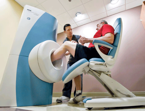
Revolutionizing Extremity Imaging: UFAI's Open MRI for the Foot and Ankle
Read More
-
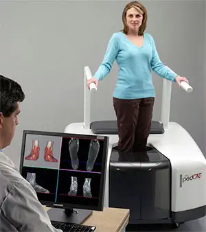 State-of-the-Art CT Scanning, Now in Our Office
Read More
-
 Listen Now
How to Choose Running Shoes: 6 Essential Steps
Read More
-
 Listen Now
15 Summer Foot Care Tips to Put Your Best Feet Forward
Read More
-
 Listen Now
Do blood pressure medicines cause foot pain?
Read More
-
 Listen Now
The Link Between Foot Health and Posture
Read More
-
 Listen Now
What is erythromelalgia?
Read More
-
 Listen Now
Flip-flops Causing You Pain? Protect Your Feet This Summer!
Read More
-
 Listen Now
Is Foot Analysis Better than Horoscopes? What Do Your Toes Reveal About Your Personality?
Read More
-
 Listen Now
Could Feet Be the Windows to Your Health?
Read More
-
 Listen Now
Why Are My Feet Different Sizes? It's More Common Than You Think
Read More
-
 Listen Now
9 Running Tips from Sports Medicine Experts
Read More
-
 Listen Now
Custom Orthotics vs. Over-the-Counter Inserts: Which Are Best for Your Feet?
Read More
-
 Listen Now
Common Foot Problems In Aging Feet: What To Watch Out For
Read More


