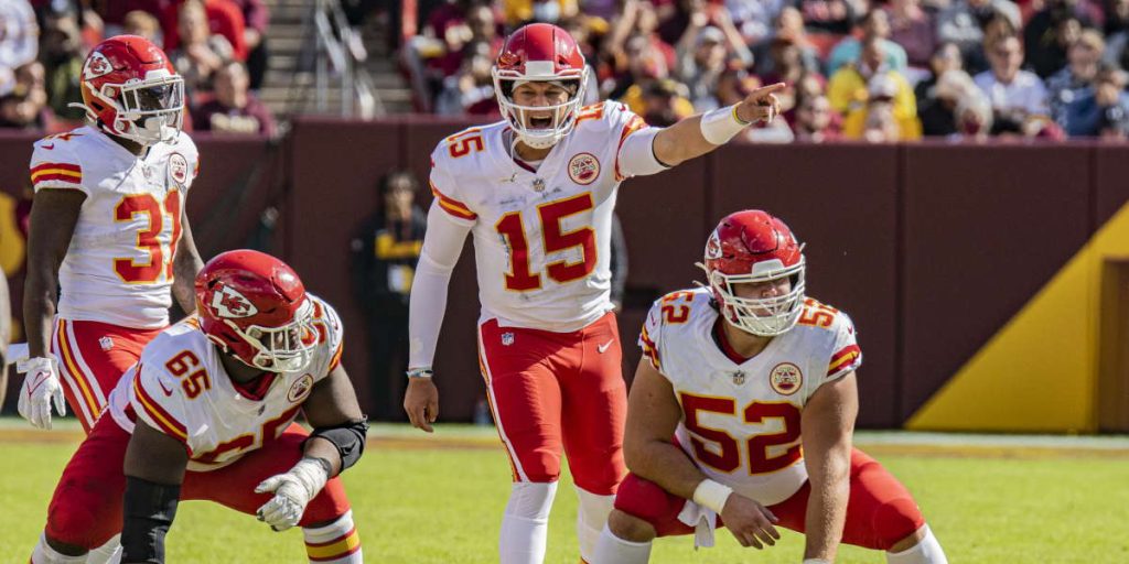
Patrick Mahomes of the Kansas City Chiefs, Sam Darnold of the Carolina Panthers, Saquon Barkley of the New York Giants, and Quandre Diggs of the Seattle Seahawks are just some of the long list of people from the NFL that recently had a high ankle sprain.
What is a high ankle sprain?
All ankle sprains are ligament injuries. Ligaments are soft tissue bands that attach bone to bone and provide stability to a joint; an injury to a ligament is called a sprain. There are three grades of injury to a ligament: grade I (mild injury), grade II (moderate injury), and grade III (severe injury).
High ankle sprains, also known as syndesmosis injuries, are common injuries in professional sports. These sprains occur when the syndesmosis (high ankle ligaments) that connect the tibia and fibula (the two lower leg bones) are stretched or torn. The syndesmosis is actually comprised of three ligaments:
- Anterior inferior tibiofibular ligament — the ligament running in front of the lower leg bones.
- Posterior inferior tibiofibular ligament — the ligament running behind the bones.
- Interosseous membrane — a supporting ligament that runs down the middle of the two bones.
High ankle sprains are commonly caused by external rotation or dorsiflexion — a sudden twist of the ankle or the foot bending up towards the shin. This is particularly common in sports that involve a lot of cutting, jumping, and changing direction such as football, soccer, basketball, and rugby. These actions put extra stress on the ankle and increase the risk of a sprained ankle.
This leads to widening between the bones at the ankle and a lack of stability. High ankle sprains often occur when a player gets rolled up on or hit from the side as their leg is firmly planted.
What sports have a high amount of high ankle sprains?
High ankle sprains can occur in any sport that involves running, jumping, or cutting maneuvers. However, some sports have a higher incidence of high ankle sprains than others. These sports include:
Basketball
A large percentage of high ankle sprains result from playing basketball. Players are at high risk of ankle injuries due to the sudden stop-and-go movements required.
Numerous NBA superstars have suffered from high ankle sprains throughout the years. Some notable examples include:
- Steph Curry of the Golden State Warriors
- Kevin Durant of the Phoenix Suns
- Joel Embiid of the Philadelphia 76ers
- Kawhi Leonard of the LA Clippers
- Paul George of the LA Clippers
- Russell Westbrook of the LA Clippers
Football, Soccer, and Rugby
These sports involve much cutting and twisting, along with high-intensity collision and contact.
These sports involve high-impact collisions and sudden changes in direction put much stress on the ankle joints. Additionally, the cleats worn by these athletes can contribute to the risk of ankle sprains as they provide less support and stability compared to regular shoes. The cleats are designed to dig into the ground for better traction, but this also means that it is less flexible and more susceptible to injury.
Volleyball
Volleyball players are at a very high risk of ankle sprains due to the repetitive jumping and landing motions required in the sport. The constant cutting and pivoting motions can also stress the ankle joints. Additionally, the surface on which volleyball is played, typically a hardwood floor, can increase the risks because it can be slippery.
It’s important to note that any sport that requires running, jumping, or cutting maneuvers, can result in a high ankle sprain. Even hiking, climbing, or running on an irregular surface can cause a high ankle sprain.
What are the symptoms of a high ankle sprain?
The symptoms of a high ankle sprain can vary depending on the severity of the injury, and some individuals may not experience all of these symptoms. The most common symptoms include:
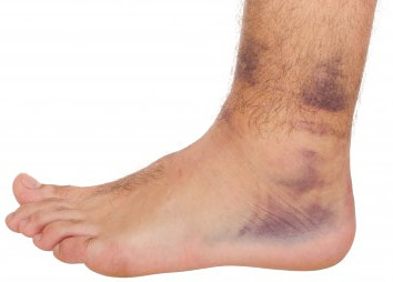
- Pain and swelling around the ankle and lower leg, particularly above the ankle joint.
- Bruising and discoloration of the skin.
- Difficulty bearing weight on the affected leg.
- Stiffness and limited range of motion in the ankle.
- Instability or a feeling of looseness in the ankle joint.
- A popping or snapping sensation at the time of injury.
- Tenderness or pain when the ankle is moved in certain positions.
- Numbness or tingling in the foot.
If you suspect you have a high ankle sprain, it’s a good idea to see a doctor for an evaluation. They may take an X-ray or CT scan to determine the extent of the injury and recommend a course of treatment.
What’s the difference between a regular ankle sprain and a high ankle sprain?
A regular ankle sprain, also known as a lateral ankle sprain or a low ankle sprain, involves stretching or tearing of the ligaments on the outside of the ankle. This type of sprain typically occurs when the foot is rolled outward, causing injury to the lateral ligaments.
A high ankle sprain involves stretching or tearing of the ligaments that connect the tibia and fibula above the ankle joint. This type of sprain typically occurs when the foot is twisted or rotated, causing injury to the syndesmotic ligaments.
Many low ankle sprains happen without contact, but a syndesmotic sprain often involves some sort of collision and a twisting motion to the lower leg.
High ankle sprains are generally considered more severe than typical ankle sprains and may take longer to heal.
How do we diagnose a high ankle sprain?
Diagnosing syndesmotic injuries is done through physical examination and radiographs. Clinically, patients with a syndesmotic ligament injury will experience pain along the ligament.
In office, a podiatrist can perform a squeeze test and an external rotation test. These tests check for pain
Imaging tests such as X-rays, CT scans, and MRIs (magnetic resonance imaging) allow podiatrists to visualize the bones and ligaments of the ankle joint. With imaging tests, a healthcare professional can diagnose an injured ankle and rule out an ankle fracture.
Although X-rays do not show ligaments, the alignment of the ankle joint, as well as associated fracture patterns of the ankle, can give signs that a syndesmotic ligament injury has occurred. In cases where the clinical and X-ray exams are inconclusive, an MRI can be performed on the ankle to visualize the ligament.
The doctor will also go over your medical history; it is important to find out if there is a history of a prior sprain or if this patient has a “trick ankle.”
If there was a “pop” or the patient could not put weight on the ankle after the injury, the odds are good that the injury was a grade II or grade III syndesmotic ankle sprain. You should be evaluated by a doctor as soon as possible.
What are the non-surgical treatments for a high ankle sprain?
Treatment for a high ankle sprain typically includes the R.I.C.E. method (Rest. Ice. Compression. Elevation.) and non-steroidal anti-inflammatory drugs (NSAIDs) to reduce pain and swelling. In some cases, an ankle brace or splint may be used to immobilize the ankle and promote healing.
Ankle braces or taping the ankle can help provide support after the patient’s return to sports. This added support can reduce risk of future injury.
Physical therapy and rehabilitation exercises may also be recommended to help strengthen the ankle and improve the range of motion.
When is surgery necessary for a high ankle sprain?
If the ankle joint is well aligned and there is no associated fracture, cast or boot immobilization for six to eight weeks is recommended for the ligament to heal.
If the ankle joint is not well aligned, and/or there is an associated displaced fracture, surgery is needed to realign the bones and ligaments. The goal of surgical correction is to realign the ankle joint. After the reduction of the ankle back into its anatomical alignment, the two methods frequently used to help stabilize the ligament are screw fixation and tightrope fixation.
What is screw fixation?
Screws have been historically used to help stabilize the syndesmotic ligament after an injury. They are placed above the ankle joint, crossing the syndesmotic ligament.
They are positional screws to help hold the tibia and fibula in place as the ligaments heal. Patients are in a non-weight bearing cast for two to three months to allow the ligaments to heal in a stable position before the screws are removed.
What is tightrope fixation?
A tightrope can also be placed across the syndesmotic ligament to help stabilize the joint. This technology is newer but provides an effective alternative to using screws to hold the ligament stable as it heals.
A tightrope is a strong suture that is held to the bone with two buttons. The buttons abut against the bone, and the suture is pulled tight, holding the bones in position as the syndesmotic ligament heals. The advantage of the tightrope is that no additional surgery is required to remove the tightrope after the syndesmotic ligament heals.
How long does it take to recover from a high ankle sprain?
The recovery time for a high ankle sprain can vary depending on the severity of the injury. According to the American Orthopaedic Foot and Ankle Society, it can take about six to eight weeks for a patient to return to normal activities. However, in more severe cases, recovery can take several months. Returning to sports or physical activity too soon can delay healing and increase the risk of re-injury.
Factors that can affect recovery time include:
- The extent of the damage to the ligaments.
- The individual’s overall health and physical condition.
- Whether or not the injury was accompanied by a fracture.
It’s important to take proper precautions to prevent these injuries and for them to receive proper treatment and rehabilitation if they do occur. With the right care, most people can fully recover and return to their normal activities.
Should I see a foot and ankle surgeon if I think I have a high ankle sprain?
It’s a good idea to see a foot and ankle surgeon if you suspect you have a high ankle sprain. High ankle sprains can be more difficult to diagnose and treat than traditional ankle sprains, and a specialist in foot and ankle care will be able to evaluate your injury and provide an appropriate treatment plan. They can also confirm the type and extent of the sprain.
A high ankle sprain can also damage the deltoid ligament which runs inside the ankle.
The goal of treatment is to prevent long-term problems, pain, or ankle instability. Rehabilitation is the key to avoiding future problems.
Why choose the University Foot & Ankle Institute for your ankle care?
Whether you need to find proper footwear, treat an injury, or get advice on proper foot care, we’re here to help. Our team of podiatrists, orthopedic surgeons, physical therapists, and sports medicine experts offers the most advanced podiatry care and the highest success rates in the nation. We are nationally recognized foot and ankle specialists and leaders in researching, diagnosing, and treating all foot and ankle conditions and common injuries.
Regular checkups with a skilled podiatrist can prevent serious foot problems by catching them early.
For a consultation please call (877) 736-6001 or make an appointment online now.
Our podiatrists take patients’ safety seriously. Our podiatry facility’s Covid-19 patient safety procedures exceed all the CDC’s coronavirus pandemic recommendations. Masks are always required in our institutes.
University Foot and Ankle Institute is conveniently located throughout Southern California and the Los Angeles area. Our foot doctors are available at locations in or near Santa Monica, Beverly Hills, West Los Angeles, Manhattan Beach, Northridge, Downtown Los Angeles, Westlake Village, Granada Hills, Santa Barbara, and Valencia.
References
Jones MH, Amendola A. Syndesmosis sprains of the ankle: a systematic review. Clin Orthop Relat Res. 2007; 455 :173–5.
Waterman BR, Belmont PJ Jr, Cameron KL, Svoboda SJ, Alitz CJ, Owens BD: Risk factors for syndesmotic and medial ankle sprain: Role of sex, sport, and level of competition. Am J Sports Med 2011;39(5):992–998.
As a teaching institution, University Foot and Ankle Institute’s Fellowship Program is among the most advanced in the nation.
We at UFAI are driven to get our patients back to their normal activities with the highest level of function, in the least amount of time, using the least invasive treatments possible. From start to finish, we are with you every step of the way.
The UFAI Education Team works to help empower our patients and website visitors with the most up-to-date information about foot and ankle conditions, treatment options, recovery and injury prevention. Our goal is to pass on truly useful information to our readers.
We hope you enjoy our work and find it of value. Please let us know!
- Could Feet Be the Windows to Your Health? - May 24, 2024
- Is Foot Analysis Better than Horoscopes? What Do Your Toes Reveal About Your Personality? - May 7, 2024
- The Link Between Foot Health and Posture - April 14, 2024